
Pathophysiology: on-target and off-target toxicity
The exact mechanism underlying ADC-related ocular adverse events is not completely understood. Several factors make the eye especially vulnerable: the abundance and variety of cell-surface receptors, large blood flow, and rapidly renewing cell subpopulations. Pathophysiology is broadly categorised into on-target and off-target toxicity, with off-target toxicity believed to be the major mechanism.
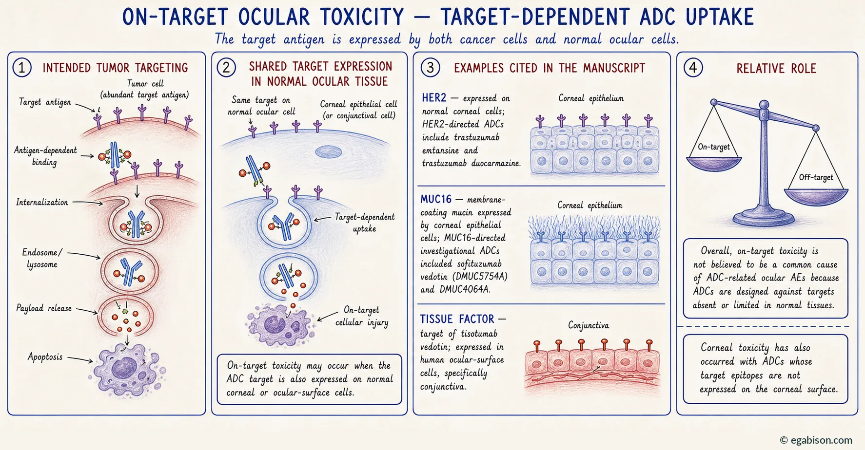
3.1. On-target toxicity
On-target toxicity involves target-dependent uptake of ADCs. It may occur when the target antigen is not only expressed on cancer cells but also on normal ocular cells.
Three examples are documented:
- HER2, expressed on normal corneal cells and targeted by trastuzumab emtansine and trastuzumab duocarmazine26.
- MUC1633, a transmembrane mucin expressed by corneal epithelial cells, targeted by the now-discontinued investigational ADCs sofituzumab vedotin (DMUC5754A) and DMUC4064A.
- Tissue factor, the target of tisotumab vedotin, expressed in human ocular surface cells, specifically the conjunctiva.
Overall, however, on-target toxicity is not believed to be a common cause of ADC-related ocular adverse events, as ADCs are developed to target sites that are not present or have limited presence in the body under normal conditions. The decisive argument is clinical: corneal toxicity has been found for ADCs targeting various epitopes not expressed on the corneal surface.
3.2. Off-target toxicity
Unlike on-target toxicity, off-target toxicity is target independent. The majority of ADC-related ocular adverse events are thought to occur through this route, with multiple contributing factors: suboptimal linker stability, receptor-mediated endocytosis, nonspecific endocytosis and the bystander effect.
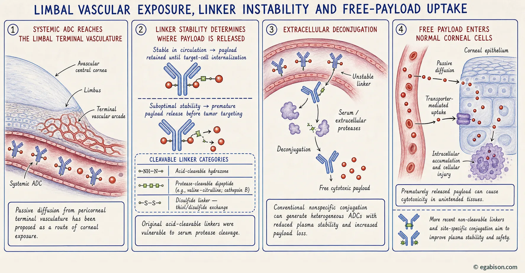
3.2.1. Linker instability and extracellular deconjugation
A linker that is insufficiently stable in circulation releases its payload before the tumour is reached. The free payload — highly lipophilic for most families in use — then enters the intracellular space of normal cells by passive diffusion or mediated transport. Conventional nonspecific conjugation compounds this, generating heterogeneous ADC populations in which the most heavily loaded fraction is the least plasma-stable.
This mechanism is particularly relevant to the cornea, whose epithelium is bathed by the tear film — a transudate of blood — and bordered at the limbus by a terminal vascular arcade. A circulating free payload therefore has two direct routes to the corneal epithelium without any antigen recognition being required.
3.2.2. Receptor-mediated endocytosis
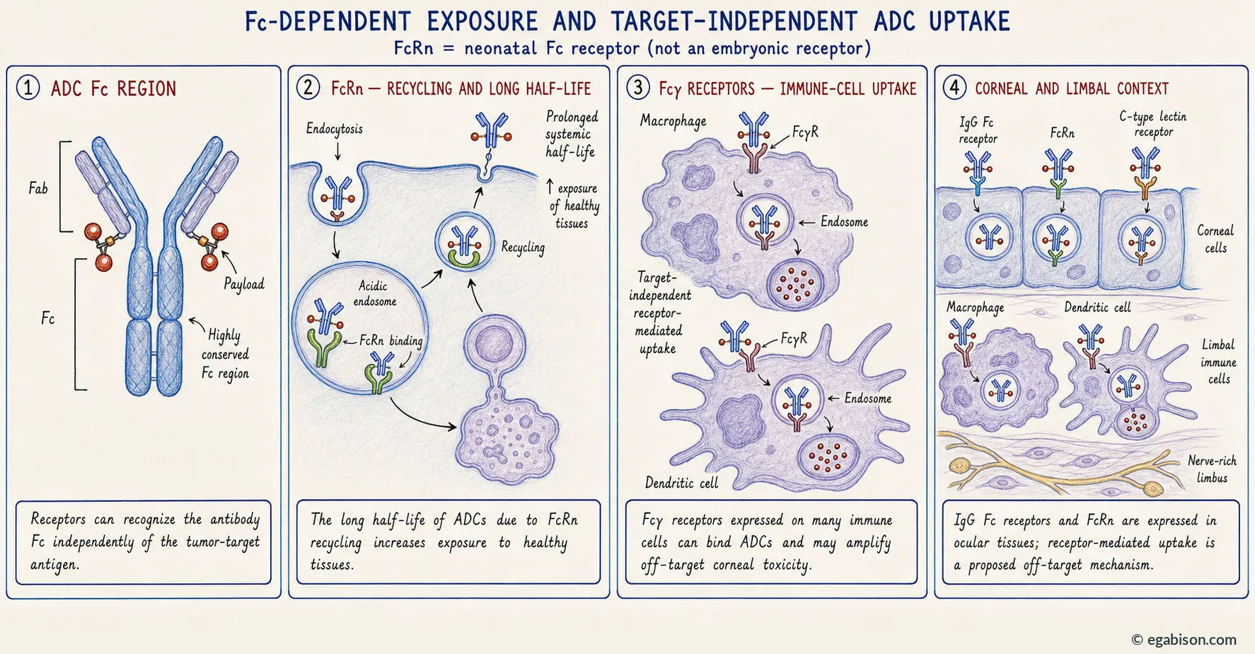
Off-target ocular adverse events may also result from target-independent receptor-mediated uptake of ADCs. Receptors including FcγRs, FcRn and C-type lectin receptors recognise and bind the highly conserved Fc regions of IgG antibodies on ADCs, potentially resulting in ADC internalisation into normal cells.
The corneal limbus region is particularly important in explaining the pathophysiology of ADCs' adverse events, due to the presence of both epithelial and stromal corneal cell progenitors, immune cells and a high nerve density. Because the IgG Fc receptor and the neonatal Fc receptor31 are expressed on the surface of corneal cells, corneal thesaurismosis could be promoted.
Two consequences compound one another. The long half-life of ADCs due to their recycling by the neonatal Fc region receptor (FcRn) increases exposure to healthy tissues; and the receptors for the Fc region of IgG (RFcγ) expressed on many cells of the immune system can bind these ADCs. Both could amplify off-target toxicity in the cornea.
The neonatal Fc receptor (FcRn) owes its name to its discovery in the context of maternal IgG transfer to the newborn. It is not an embryonic receptor and remains ubiquitously expressed in adulthood, where it recycles IgG and accounts for their prolonged half-life. It is this function, not any neonatal specificity, that operates in ocular ADC exposure.
3.2.3. Nonspecific endocytosis: macro- and micropinocytosis
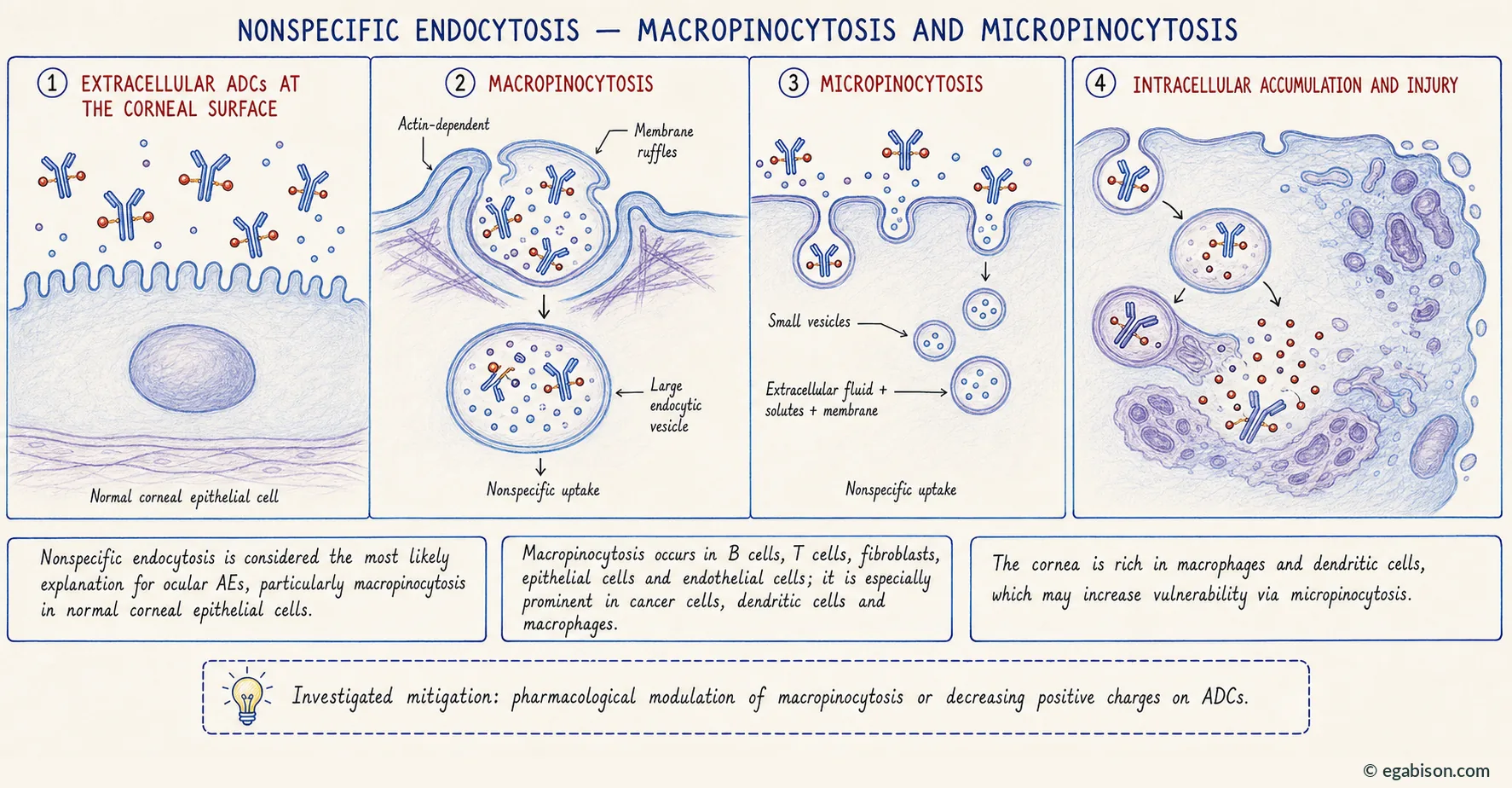
Nonspecific endocytosis — including macropinocytosis and micropinocytosis, the latter being nonspecific internalisation of very small vesicles containing extracellular fluid, solutes and membrane — is the most likely explanation for ocular adverse events, particularly macropinocytosis, due to ADC uptake by normal corneal epithelial cells.
Macropinocytosis is the nonspecific, actin-dependent internalisation of large amounts of extracellular fluid, solutes and membrane in large endocytic vesicles. It involves the development of ruffled extensions of the plasma membrane in areas of abundant extracellular fluid to facilitate endocytosis.
This mechanism occurs in various cell types — B and T cells, fibroblasts, epithelial cells and endothelial cells — but is especially prominent in cancer cells, dendritic cells and macrophages, where micropinocytosis is a constitutive process. The cornea is rich in macrophages and dendritic cells, which may make it especially vulnerable to off-target toxicity via micropinocytosis.
This route has a direct therapeutic implication: it is modulable. To minimise these adverse events, modulation of macropinocytosis29 is currently being investigated through pharmacological modulation of the cellular process or by decreasing positive charges present on ADCs.
3.2.4. Bystander effect
Off-target toxicity can also result from a bystander effect, a phenomenon that occurs when, after reaching its target, free payload released from antigen-positive target cells travels to the extracellular space — due to permeability of a cell, high lipophilicity of the payloads, or diminished cell integrity following cell death — and then enters the intracellular space of antigen-negative normal cells via passive diffusion, transporter-mediated uptake or nonspecific endocytosis, causing damage of the noncancerous surrounding cells.
This effect, sought after for antitumour purposes so as to reach low-antigen-expressing tumour cells within a heterogeneous tumour, is inseparable from the risk of neighbouring-tissue toxicity. It exemplifies a property whose benefit and risk arise from the same physicochemical determinant — payload lipophilicity.
Why the cornea and the limbus?
The question deserves separate treatment, because it governs the understanding of the clinical phenotypes. Most ADC-related ocular adverse events are corneal in nature. Corneal toxicity has been found for ADCs targeting various epitopes not expressed on the corneal surface. The reason for the corneal distribution of these ADCs has been the subject of hypotheses, including the bathing of corneal epithelial cells by tears, which are a transudate of blood, and simple passive diffusion from the peri-corneal terminal vasculature.
A reduction in blood flow to this terminal vascularisation has been tested by both physical means — application of cold — and pharmacological means — vasoconstrictors applied to the ocular surface throughout the treatment or simply during the period surrounding the infusions. The rationale for these measures follows directly from the vascular hypothesis.
The corneal limbus region is particularly important in explaining the pathophysiology of ADCs' adverse events, due to the presence of both epithelial and stromal corneal cell progenitors, immune cells and a high nerve density. Four features converge there:
- a stem cell niche in permanent renewal, whose transient amplifying cells are by definition actively dividing — hence elective targets for a microtubule inhibitor;
- a terminal vascular arcade that delivers any circulating molecule locally;
- a resident immune population — macrophages and dendritic cells — that is constitutively pinocytotic;
- a nerve density among the highest in the body, exposing the subbasal plexus to direct neurotoxicity.
This convergence explains why ADC corneal toxicity is not limited to a diffuse toxic keratitis but adopts characteristic topographies — peripheral first, centripetal thereafter — that betray its limbal origin.