
Pathological corneal healing: direct epithelial–stromal interactions
Original teaching synthesis, based on the review by Gabison EE, Huet E, Baudouin C, Menashi S. "Direct epithelial–stromal interaction in corneal wound healing: role of EMMPRIN/CD147 in MMPs induction and beyond", Progress in Retinal and Eye Research 2009;28:19-33, and on the original work of Pr Éric Gabison's team.
The transparent cornea owes its clarity to an architecture of strictly separated compartments: a surface epithelium, a stroma that makes up most of its thickness, and membranes that isolate them from one another. This separation is not an anatomical detail — it determines the outcome of every episode of corneal wound healing. When an injury durably breaks it down, the epithelium and stromal cells come into direct contact, an event that is always pathological and that triggers a dysregulated proteolytic cascade [1].
This course traces, over three parts, the mechanism of direct epithelial–stromal interactions (direct-ESI): the physiology of the barriers that normally prevent them, the molecular loop — centered on the glycoprotein EMMPRIN/CD147 — that amplifies them once triggered, and the two healing outcomes, fibrosis or stromal melting, that they determine.
An architecture of separate compartments
The cornea is made of three distinct layers — epithelium, stroma, endothelium — themselves separated by dedicated structures: the epithelial basement membrane and Bowman's layer between epithelium and stroma, and Descemet's membrane between stroma and endothelium. This compartmentalization is considered essential to corneal physiology, and abnormalities in the regeneration of these membranes after injury are associated with pathological forms of wound healing, from subepithelial haze (fibrosis) to ulceration and perforation [1].
Exchanges between epithelium and stroma normally occur through diffusible cytokines — IL-1, TGF-β, PDGF, KGF, HGF — which provide bidirectional communication at a distance, without cell contact. It is when this distant signaling is replaced by direct cell–cell contact that the epithelial–stromal dialogue shifts into a pathological mode [1].
Physiological epithelial–stromal barriers
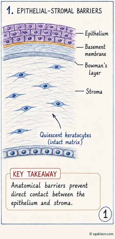
A selectively permeable basement membrane
The basement membrane is not a simple inert partition: it exerts selective permeability toward the cytokines produced by the epithelium. Under homeostatic conditions, IL-1, TGF-β and PDGF remain sequestered in the epithelial compartment — partly due to their affinity for heparan sulfate in the basement membrane — and only reach the stroma after this barrier is disrupted. Conversely, other factors such as KGF can exert their biological effects through an intact basement membrane [1].
The role of this membrane goes beyond a simple physical barrier: its maturation actively inhibits TGF-β production by the regenerating epithelium, an antifibrotic protection mechanism documented by lower tear TGF-β levels after LASEK (which preserves the basement membrane) than after PRK [1].
Bowman's layer, a second line of defense
Beneath the epithelial basement membrane, Bowman's layer — an acellular band, 8 to 15 µm thick, made of extracellular matrix — constitutes a second barrier against direct epithelial–stromal contact. Its precise biological role remains debated: some authors consider it non-essential, pointing to the relative safety of PRK, which sections it without regenerating it; others attribute to it a role in biomechanical rigidity and corneal curvature. But unlike the epithelial basement membrane, Bowman's layer does not regenerate once destroyed — making its loss, in the context of direct-ESI, a potentially permanent one [1].
Two successive anatomical barriers — the epithelial basement membrane (regenerable) and Bowman's layer (non-regenerable) — normally protect the stroma from direct contact with the epithelium. Their integrity determines the mode of wound healing.
Injury, persistent epithelial defect & barrier breakdown
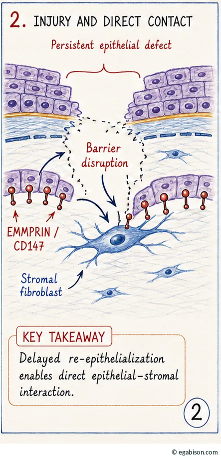
From delayed healing to persistent epithelial defect
A common triggering event across most corneal ulcerations, whatever their cause — tear film abnormality, toxic reaction, neurotrophic or metabolic disorder — is a delay in epithelial closure. When an epithelial defect fails to heal within about one week, it is termed a persistent epithelial defect (PED) [1,8]. Predisposing conditions include neurotrophic keratitis (see the dedicated course), severe dry eye, limbal stem cell deficiency and sequelae of chemical burns.
Basement membrane dissolution and stromal exposure
In the absence of closure, a PED progresses toward basement membrane dissolution — a prerequisite for stromal thinning [5,6]. After the initial injury, subepithelial keratocytes first undergo apoptosis, creating an acellular zone between epithelium and stroma; in normal healing, this zone closes as the basement membrane is reconstituted, which keeps keratocytes separated from the regenerating epithelium. But a prolonged delay slows this maturation, sometimes as far as the disappearance of Bowman's layer — which does not regenerate —, and stromal repopulation then ends up occurring in the absence of a barrier, resulting in direct, potentially lasting contact between epithelium and keratocytes: these are the direct epithelial–stromal interactions (direct-ESI), always pathological [1].
An epithelial defect that persists beyond one to ten days should never be regarded as simple slowness: it is a warning sign of an imminent risk of direct-ESI and stromal melting, requiring active investigation of the cause and intervention before direct contact becomes established.
Glossary of abbreviations used in this course
Scientific acronyms and abbreviations used across the 3 pages of this course, listed alphabetically.
No matching term.
- AMT
- Amniotic Membrane Transplantation
- α-SMA
- alpha-smooth muscle actin, myofibroblast marker
- cDNA
- complementary DNA, used in transfection to overexpress a protein
- CD147
- cluster of differentiation 147; synonym of EMMPRIN and basigin
- ECM
- extracellular matrix
- EGF
- Epidermal Growth Factor
- EMMPRIN
- Extracellular Matrix Metalloproteinase Inducer; synonym of CD147
- FAK
- Focal Adhesion Kinase
- direct-ESI
- direct epithelial–stromal interactions
- Ig
- immunoglobulin domain, extracellular structural motif of EMMPRIN
- IL-1
- interleukin-1
- KC
- keratoconus
- LASEK
- Laser-Assisted Sub-Epithelial Keratomileusis
- LASIK
- Laser-Assisted In Situ Keratomileusis
- MMP
- Matrix Metalloproteinase
- PDGF
- Platelet-Derived Growth Factor
- PED
- persistent epithelial defect
- PRK
- photorefractive keratectomy (surface laser photoablation)
- siRNA
- small interfering RNA
- Smad
- effector proteins of the TGF-β signaling pathway
- TGF-β
- Transforming Growth Factor beta
- TIMP
- Tissue Inhibitor of Metalloproteinases
- uPA
- urokinase-type Plasminogen Activator