
Two healing outcomes
Two opposing myofibroblast phenotypes
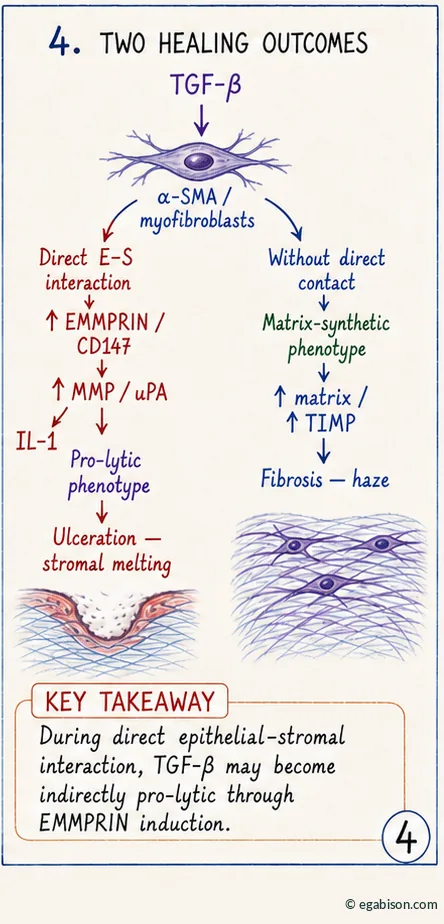
Diffusible signaling: the "fibrotic" myofibroblast
When TGF-β acts through diffusible signaling, without direct contact, it outweighs the effect of IL-1, inhibits collagenase expression and increases expression of their inhibitor, TIMP-1. The fibroblast that differentiates under these conditions adopts a synthetic phenotype: matrix accumulation, contraction, and ultimately subepithelial fibrosis (haze) — the classic mode of wound healing associated with TGF-β.
Direct contact mediated by EMMPRIN: the "pro-lytic" myofibroblast
At direct epithelial–stromal contact, the picture reverses. EMMPRIN expression remains under the transcriptional control of TGF-β, but the function of TGF-β then flips: from anti-proteolytic in diffusible signaling, it becomes pro-lytic by inducing the EMMPRIN → MMP/uPA axis. The result is a second myofibroblast phenotype, just as α-SMA-positive as the first but functionally opposite — a producer of MMPs rather than matrix — preferentially found in ulcerated corneas, where matrix degradation dominates [1,4].
A single marker, α-SMA, can correspond to two functionally opposite myofibroblast phenotypes. It is not the presence of TGF-β that determines the healing outcome, but its route of induction — diffusible (fibrosis) or mediated by direct contact through EMMPRIN (melting).
Natural protective mechanisms against direct-ESI
Anatomical barriers: basement membrane and Bowman's layer
The cornea has natural defense mechanisms against direct-ESI, which explains the relative rarity of ulcerations and perforations given the constant environmental aggressions it endures. The strict compartmentalization of its layers, ensured by the epithelial basement membrane and Bowman's layer (see chapter 2), forms the first line of defense — this is, in fact, the documented protective role of amniotic membrane transplantation, which accelerates epithelial closure while providing a substitute barrier against direct contact [1].
Keratocyte apoptosis: a dynamic barrier
Alongside these structural barriers, a dynamic mechanism operates: after an epithelial injury, the underlying keratocytes undergo apoptosis, creating a transient acellular zone between epithelium and stroma. Epithelial repair consistently precedes stromal repopulation by fibroblasts, so this asynchrony naturally delays any contact — for as long as it takes the basement membrane to reconstitute. It is only when epithelial closure is delayed that fibroblasts regain the anterior stroma before the barrier is restored, allowing direct contact to occur [1].
Direct-ESI therefore do not arise from a deep lesion alone, however severe: it is the delay in epithelial healing that, by leaving barriers disrupted for longer and giving fibroblasts time to recolonize an unprotected stroma, turns an ordinary injury into a pathological interaction.
Clinical settings, therapeutic implications & references
Where do direct-ESI occur clinically?
Beyond "classic" ulcerations — rheumatoid arthritis, Sjögren syndrome, alkali burn, iatrogenic post-surgical melts — several clinical situations share this same mechanism of prolonged barrier breakdown:
- Epithelial ingrowth after LASIK: invasion of surface epithelial cells under the corneal flap; usually mild and self-limiting, but in severe cases persistent ingrowth allowing prolonged direct contact can lead to flap melt.
- Radial keratotomy: deep stromal incisions can trap epithelial plugs, with abnormal basement membrane and absent Bowman's layer regeneration, favoring long-term wound-healing defects.
- Keratoconus: rupture of the basement membrane and Bowman's layer is an early and consistent feature, associated with increased protease activity; EMMPRIN levels are increased in the stroma, particularly at sites of BM/Bowman rupture, making it a candidate mechanism in the MMP induction observed in this dystrophy.
Targeting EMMPRIN: a therapeutic strategy
Direct MMP inhibition faces a significant obstacle: these enzymes also fulfill essential physiological functions in normal wound healing — in MMP-9-deficient mice, healing is indeed accelerated, but stromal transparency is not fully restored. Targeting EMMPRIN upstream offers a double advantage: it specifically acts on the MMPs induced by the pathological direct-ESI mechanism, and it intervenes before their secretion and activation, unlike classic synthetic inhibitors which only act afterward [1]. Pending EMMPRIN-specific therapies, tetracyclines (doxycycline), already used clinically for their anti-collagenolytic effect independent of their antibacterial action, remain the closest available option to this principle (see the review of pharmacological modulators of corneal wound healing).
Summary & key points
- The cornea owes its transparency to a strict compartmentalization between epithelium and stroma, maintained by the epithelial basement membrane (regenerable) and Bowman's layer (non-regenerable).
- A persistent epithelial defect (> 1 week) exposes the cornea to dissolution of these barriers and direct contact between epithelium and keratocytes — the direct epithelial–stromal interactions (direct-ESI), always pathological.
- EMMPRIN/CD147, a transmembrane glycoprotein, mediates this contact and induces MMP-1, -2, -3 and uPA in fibroblasts, with a self-amplification loop that propagates proteolysis.
- Depending on the route of TGF-β induction — diffusible or via EMMPRIN-mediated direct contact — the myofibroblast adopts a synthetic (fibrosis) or pro-lytic (stromal melting) phenotype.
- Natural mechanisms — anatomical barriers and dynamic keratocyte apoptosis — normally protect against direct-ESI; it is delayed healing that defeats them.
- Targeting EMMPRIN upstream of MMPs is a promising therapeutic strategy, specific to the pathological mechanism and without compromising the physiological functions of MMPs.
It is not the depth of the initial injury that determines the outcome, but the time during which epithelium and stroma remain in direct contact. Preventing or shortening direct-ESI — by accelerating re-epithelialization — remains the most robust therapeutic lever against stromal melting.
Original teaching synthesis. Main source: Gabison EE, Huet E, Baudouin C, Menashi S. "Direct epithelial–stromal interaction in corneal wound healing: role of EMMPRIN/CD147 in MMPs induction and beyond", Progress in Retinal and Eye Research 2009;28(1):19-33.
Main source
- Gabison EE, Huet E, Baudouin C, Menashi S. Direct epithelial–stromal interaction in corneal wound healing: role of EMMPRIN/CD147 in MMPs induction and beyond. Prog Retin Eye Res 2009;28(1):19-33. doi:10.1016/j.preteyeres.2008.11.001.
Additional references cited in this course
- Gabison EE, Mourah S, Steinfels E, et al. Differential expression of extracellular matrix metalloproteinase inducer (CD147) in normal and ulcerated corneas: role in epithelio-stromal interactions and matrix metalloproteinase induction. Am J Pathol 2005;166(1):209-219. doi:10.1016/S0002-9440(10)62245-6.
- Huet E, Vallée B, Delbé J, et al. EMMPRIN modulates epithelial barrier function through a MMP-mediated occludin cleavage: implications in dry eye disease. Am J Pathol 2011;179(3):1278-1286. doi:10.1016/j.ajpath.2011.05.036.
- Huet E, Gabison EE, Mourah S, Menashi S. Role of EMMPRIN/CD147 in tissue remodeling. Connect Tissue Res 2008;49(3):175-179.
- Matsubara M, Zieske JD, Fini ME. Mechanism of basement membrane dissolution preceding corneal ulceration. Invest Ophthalmol Vis Sci 1991;32(13):3221-3237.
- Fini ME, Parks WC, Rinehart WB, et al. Role of matrix metalloproteinases in failure to re-epithelialize after corneal injury. Am J Pathol 1996;149(4):1287-1302.
- Cavaillé M, Tolosa Leal C, Gabison E. Cicatrisation cornéenne (chapter, collective ophthalmology textbook).
- Ma JJ, Dohlman CH. Mechanisms of corneal ulceration. Ophthalmol Clin North Am 2002;15(1):27-33.
Glossary of abbreviations used in this course
Scientific acronyms and abbreviations used across the 3 pages of this course, listed alphabetically.
No matching term.
- AMT
- Amniotic Membrane Transplantation
- α-SMA
- alpha-smooth muscle actin, myofibroblast marker
- cDNA
- complementary DNA, used in transfection to overexpress a protein
- CD147
- cluster of differentiation 147; synonym of EMMPRIN and basigin
- ECM
- extracellular matrix
- EGF
- Epidermal Growth Factor
- EMMPRIN
- Extracellular Matrix Metalloproteinase Inducer; synonym of CD147
- FAK
- Focal Adhesion Kinase
- direct-ESI
- direct epithelial–stromal interactions
- Ig
- immunoglobulin domain, extracellular structural motif of EMMPRIN
- IL-1
- interleukin-1
- KC
- keratoconus
- LASEK
- Laser-Assisted Sub-Epithelial Keratomileusis
- LASIK
- Laser-Assisted In Situ Keratomileusis
- MMP
- Matrix Metalloproteinase
- PDGF
- Platelet-Derived Growth Factor
- PED
- persistent epithelial defect
- PRK
- photorefractive keratectomy (surface laser photoablation)
- siRNA
- small interfering RNA
- Smad
- effector proteins of the TGF-β signaling pathway
- TGF-β
- Transforming Growth Factor beta
- TIMP
- Tissue Inhibitor of Metalloproteinases
- uPA
- urokinase-type Plasminogen Activator