
Corneal macrophages: anatomy & immune privilege
Original teaching synthesis based on the review by Yaghmour A, Arabpour Z, Al-Khudari H, Djalilian A. "When Macrophages Heal and When They Scar: Timing in Corneal Fibrosis", Life 2026;16:1090 (open access), illustrated by Pr Éric Gabison's team.
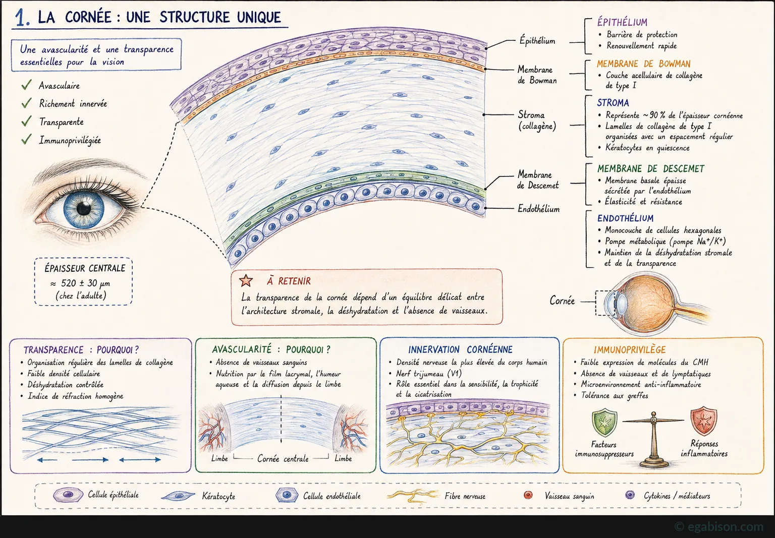
The cornea is avascular but not devoid of immune cells: macrophages reside there permanently, at low density, mainly at the periphery and limbus, without compromising transparency or avascularity [Yaghmour 2026]. Their role is to survey the tissue, orchestrate defence against injury, and then close the inflammatory window at the right time to allow transparent healing.
This immune privilege rests on an active balance, not a simple absence: low expression of MHC molecules, absence of blood and lymphatic vessels, an anti-inflammatory microenvironment (soluble immunosuppressive factors, TGF-β, α-MSH, ACAID) and relative immune tolerance to grafts. Disrupting this balance — through trauma, infection or repeated surgery — exposes the cornea to dysregulated inflammation and fibrosis [Streilein 2003].
Resident vs recruited populations
Two macrophage populations coexist in the cornea, with non-redundant and temporally distinct roles [Yaghmour 2026]:
- Resident macrophages: of embryonic origin, they self-renew in place. They provide immune surveillance, maintain immune privilege, clear debris and drive early resolution of minor injuries. They are especially important for preserving stromal quiescence.
- Monocyte-derived macrophages: rapidly recruited from the circulation via limbal vessels in response to epithelial injury, chemokine gradients and vascular leakage. They dominate during acute, severe injury and are the main source of the cytokines, growth factors and matrix-modifying enzymes that drive early defence and stromal remodelling.
Corneal fibrosis is not simply a consequence of macrophage presence: it reflects a failure to transition from recruited inflammatory programmes toward resident-like or resolution-phase states. The therapeutic target is therefore not to eliminate macrophages, but to restore their proper timing [Yaghmour 2026].
Timeline of macrophage responses
The macrophage response to corneal injury follows a dynamic four-phase continuum, which determines the outcome of healing — transparent regeneration or fibrosis [Yaghmour 2026].
Four phases, one clock
| Phase | Time | Goal | Possible outcome |
|---|---|---|---|
| Early inflammatory | 0 h – 1 to 3 days | Host defence and debris clearance | Infection control, initiation of repair |
| Early repair | 3 to 7 days | Promotion of tissue repair | Effective repair |
| Resolution | > 7 days | Resolution of inflammation and restoration | Restoration of functional transparency |
| Chronic dysregulated | Weeks to months | Persistent inflammation and matrix deposition | Fibrosis, opacification, loss of transparency |
Each phase is dominated by a distinct macrophage phenotype: the pro-inflammatory (M1-like) macrophage phagocytoses pathogens and debris and secretes IL-1β, TNF-α, IL-6, ROS and NO; intermediate states secrete growth factors (VEGF, PDGF) and initiate matrix remodelling; the pro-resolving (M2-like) macrophage clears apoptotic cells by efferocytosis and secretes IL-10 and low-dose TGF-β; if this transition fails, a persistent pro-fibrotic macrophage maintains sustained secretion of TGF-β, CTGF and PDGF, chronically activating myofibroblasts.
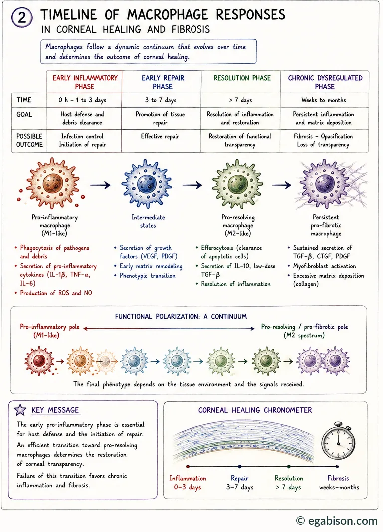
The early pro-inflammatory phase is essential for host defence and the initiation of repair. An efficient transition toward pro-resolving macrophages determines the restoration of corneal transparency. Failure of this transition favours chronic inflammation and fibrosis.
When the macrophage turns rogue
Macrophages become pathogenic drivers of fibrosis when their inflammatory activation persists beyond the appropriate temporal window. In severe chemical injury, chronic infection, autoimmune disease or repeated surgical trauma, they maintain a pro-inflammatory and pro-fibrotic secretory profile well beyond the expected proliferative phase [Yaghmour 2026]. The cytokines and growth factors they then release reinforce fibroblast-to-myofibroblast differentiation, sustain excess extracellular matrix production and prevent resolution. They also indirectly promote angiogenesis and lymphangiogenesis — two breaches of immune privilege that perpetuate inflammatory cell influx and shift healing from a regenerative to a pathological mode.
An eye that remains red and inflamed beyond the first week after corneal injury should not be read as simple slow healing: it is the warning sign of persistent pro-fibrotic macrophage activity, to be corrected before it locks in permanent opacity.
Glossary of abbreviations used in this course
Scientific abbreviations and acronyms used throughout the 10 pages of this course, listed alphabetically.
No matching term.
- 95% CI
- 95% confidence interval
- AAV
- adeno-associated virus (gene-therapy viral vector)
- ABCG2
- ABCG2 transporter, a limbal stem-cell marker
- AMT
- amniotic membrane transplantation
- anti-VEGF
- anti-angiogenic treatment targeting VEGF
- ASC
- inflammasome adaptor protein (Apoptosis-associated Speck-like protein containing a CARD)
- BrdU
- bromodeoxyuridine, a cell-proliferation marker
- CCL2
- CCL2 chemokine, synonym of MCP-1; recruits circulating monocytes
- CCR2
- receptor for CCL2; marker of newly recruited monocytes/macrophages
- CD147
- cluster of differentiation 147; synonym of EMMPRIN and basigin
- CD163
- M2 macrophage marker; scavenger receptor for haemoglobin-haptoglobin
- CD206
- mannose receptor; marker of M2 polarization
- CD74
- cell-surface receptor for MIF
- CDVA
- corrected distance visual acuity
- CGRP
- calcitonin gene-related peptide
- CTGF
- connective tissue growth factor
- CX3CL1
- fractalkine, a chemokine involved in monocyte/macrophage recruitment
- CXCR2/CXCR4
- chemokine receptors, also receptors for MIF
- DAMP
- damage-associated molecular pattern
- DESI
- direct epithelial–stromal interaction
- EGF
- epidermal growth factor
- EGFR
- epidermal growth factor receptor
- EMMPRIN
- extracellular matrix metalloproteinase inducer; synonym of CD147
- ETDRS
- reference visual-acuity scale used in clinical research (Early Treatment Diabetic Retinopathy Study)
- FasL
- Fas ligand, an apoptosis-inducing protein
- FISH
- fluorescence in situ hybridisation
- GAG
- glycosaminoglycan(s)
- GM-CSF
- granulocyte-macrophage colony-stimulating factor
- GVHD
- graft-versus-host disease
- HGF
- hepatocyte growth factor
- HIF-3α
- hypoxia-inducible factor, 3-alpha subunit
- HMGB1
- high mobility group box 1, an alarmin released by injured cells (DAMP)
- IFN-γ
- interferon gamma
- IGF-1
- insulin-like growth factor 1
- IL-1
- interleukin-1
- iNOS
- inducible nitric oxide synthase
- IPAS
- inhibitory PAS domain protein
- KGF
- keratinocyte growth factor
- LASIK
- laser-assisted in situ keratomileusis
- LogMAR
- logarithmic visual-acuity scale
- LOX
- lysyl oxidase, a collagen cross-linking enzyme
- LPS
- bacterial lipopolysaccharide
- M1
- classically activated macrophage, pro-inflammatory profile
- M2
- alternatively activated macrophage, pro-resolving/reparative profile (subtypes M2a-M2d, M2eff)
- MEN2B
- multiple endocrine neoplasia type 2B
- MerTK
- receptor tyrosine kinase involved in efferocytosis
- MIF
- macrophage migration inhibitory factor
- MRI
- magnetic resonance imaging
- NGF
- nerve growth factor
- NK
- neurotrophic keratitis
- NLRP3
- NLRP3 inflammasome (NOD-, LRP- and pyrin domain-containing protein 3)
- NO
- nitric oxide
- NSAID
- nonsteroidal anti-inflammatory drug
- PACAP
- pituitary adenylate cyclase-activating polypeptide
- PAF
- platelet-activating factor
- PAMP
- pathogen-associated molecular pattern
- PDGF
- platelet-derived growth factor
- PED
- persistent epithelial defect
- PEDF
- pigment epithelium-derived factor
- PGE₂
- prostaglandin E2
- PRK
- photorefractive keratectomy (laser surface ablation)
- pro-NGF
- precursor form of NGF
- RGTA
- matrix-therapy ReGeneraTing Agent, e.g. Cacicol
- rhNGF
- recombinant human NGF (cenegermin)
- ROS
- reactive oxygen species
- Smad
- signalling proteins of the TGF-β pathway
- SPK
- superficial punctate keratitis
- sVEGFR-1
- soluble form of VEGF receptor-1 (= sFlt-1)
- sVEGFR-3
- soluble form of VEGF receptor-3
- TGF-β
- transforming growth factor beta
- TIMP
- tissue inhibitor of metalloproteinases
- TLR
- Toll-like receptor
- TNF-α
- tumour necrosis factor alpha
- TRPV4
- transient receptor potential vanilloid 4, a mechanosensitive ion channel
- uPA
- urokinase-type plasminogen activator
- V1
- ophthalmic branch of the trigeminal nerve
- VEGF
- vascular endothelial growth factor
- VEGF-C
- lymphangiogenic isoform of VEGF
- VIP
- vasoactive intestinal peptide
- YAP/TAZ
- Hippo pathway effectors, mechanosensors of matrix stiffness
- α-SMA
- alpha-smooth muscle actin, a myofibroblast marker