
Ocular toxicity of antibody-drug conjugates
Antibody-drug conjugates (ADCs) occupy an expanding place in the therapeutic armamentarium for solid and haematological malignancies. Their principle rests on the covalent coupling of a highly potent cytotoxic payload to a monoclonal antibody directed against an antigen preferentially expressed by tumour cells. The objective is to maximise cancer cell death while minimising cytotoxicity toward noncancerous cells.
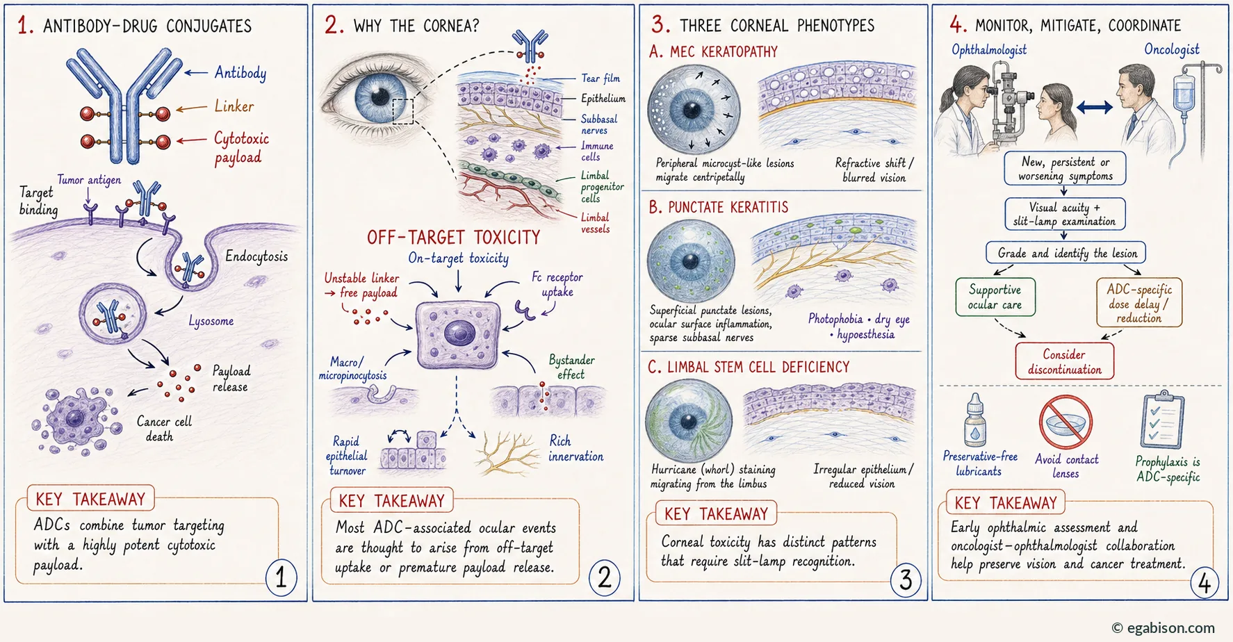
This promise of a widened therapeutic index has not translated into an absence of organ toxicity. Across phase I to III programmes, the ocular surface has emerged as one of the most frequently reported sites of dose-limiting toxicity for several ADC families. Reported manifestations are dominated by dry eye, conjunctivitis, photophobia, blurred vision and a spectrum of corneal abnormalities, of which microcyst-like epithelial keratopathy is the most characteristic signature.
Ocular toxicity of anticancer treatment is not new. It is well described for conventional cytotoxics such as cytarabine and docetaxel, with severity ranging from asymptomatic findings to vision-threatening disease. Newer targeted agents — immune checkpoint inhibitors, MEK inhibitors, tyrosine kinase inhibitors, FGFR inhibitors, BRAF inhibitors and EGFR inhibitors — have broadened this spectrum. ADCs fall within this continuum, with one distinctive feature: the frequency and early onset of ocular surface involvement are such that, for certain agents, they govern the conduct of the anticancer treatment itself.
The stakes extend beyond patient comfort. Ocular symptoms impair health-related quality of life and cause interruption, dose reduction and occasionally permanent discontinuation of anticancer therapy, with a demonstrated impact on disease control. Early recognition, accurate grading and appropriate management of these events are therefore components of the oncology pathway rather than a mere ophthalmic corollary.
This chapter restates, develops and illustrates the content of the invited review published in Progress in Retinal and Eye Research. It is built around original plates designed to render visible the proposed mechanisms, the corneal phenotypes and the management algorithms. The pathophysiology presented remains, for the most part, hypothetical: the mechanisms described are those that the literature and clinical experience render most plausible, and are labelled as such.
ADC architecture and pharmacology
2.1. The three components
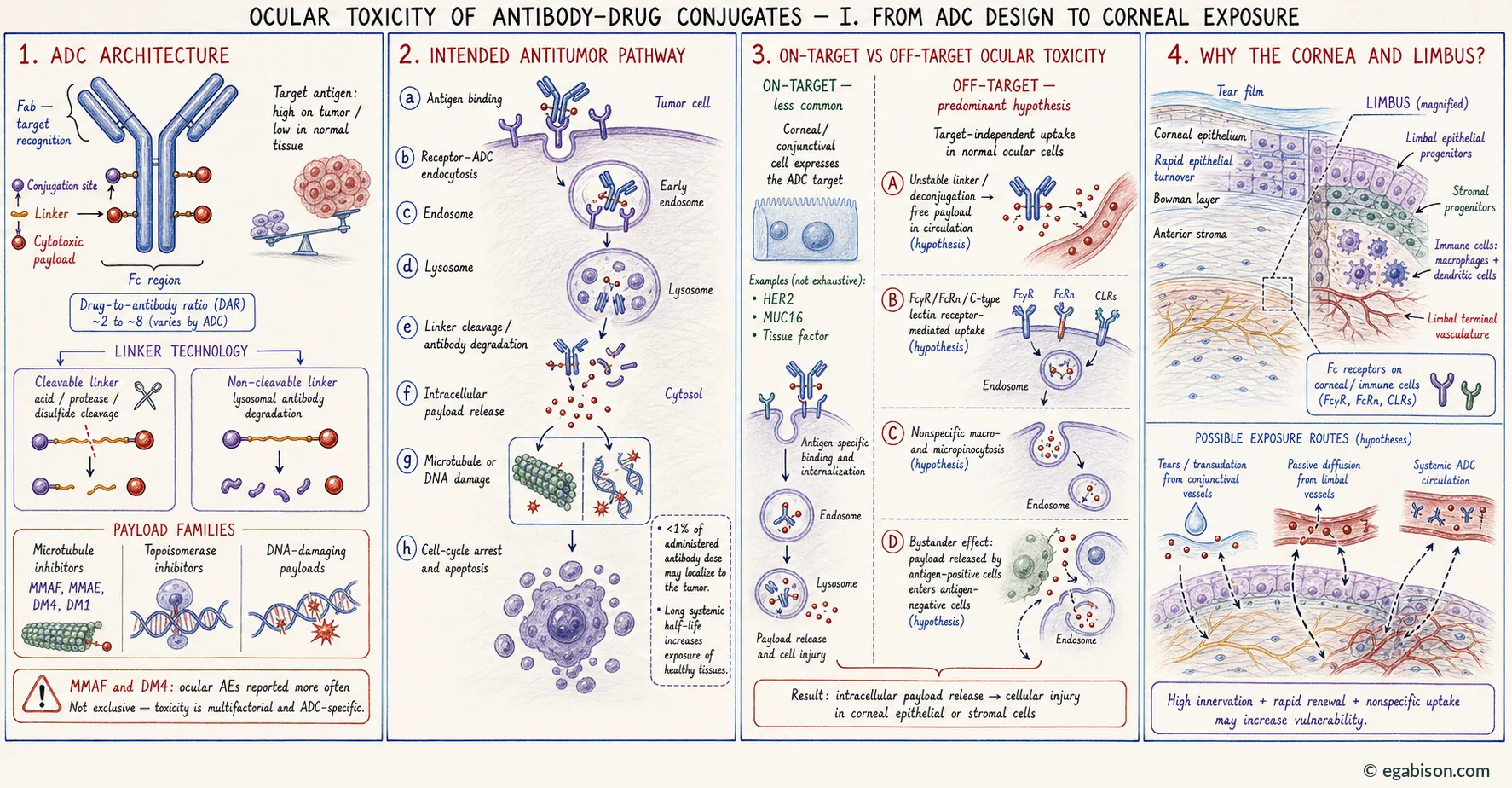
An ADC comprises three inseparable elements, each of which conditions both efficacy and safety: the monoclonal antibody, the cytotoxic payload — also known as the warhead — and the linker joining them.
For an optimised ratio between efficacy and safety, the antibody needs to bind specifically and effectively to its target antigen on cancer cells while avoiding normal non-cancer cells, to minimise the risk of adverse events. Most antibodies used in ADCs are IgG1 or IgG4 immunoglobulins designed to target tumour-specific sites. The drug-to-antibody ratio (DAR) ranges approximately from 2 to 8 depending on the agent, and is a key determinant of tissue exposure.
The cytotoxic payload exerts its effects on cellular processes required for survival. Payloads are often cytotoxic in the picomolar concentration range, which is crucial because only a very small amount — less than 1 % — of the administered antibody dose localises to the tumour, owing to limited penetration of the antibody into the tumour and poor linker stability. The remaining antibody dose is systemically released and thereby becomes available to healthy tissues.
2.2. Payload families
Three main payload families dominate the current landscape:
- Microtubule inhibitors: auristatins (MMAE, MMAF) and maytansinoids (DM1, DM4, ravtansine, mertansine), which disrupt microtubule dynamics, mitosis and intracellular transport.
- Topoisomerase I inhibitors: exatecan derivatives (deruxtecan), SN-38, which inhibit DNA replication and induce apoptosis.
- DNA-damaging agents: calicheamicin derivatives, pyrrolobenzodiazepine (PBD) dimers and duocarmycin-type alkylating agents.
The increased risk of ocular adverse events appears related to the cytotoxic payload: ocular events are more common with ADCs containing the microtubule inhibitors DM4 (a maytansine derivative) or monomethyl auristatin F (MMAF). It is unclear whether the eye is particularly sensitive to these payloads, or whether their toxicity is simply more readily detected there.
Three factors argue for enhanced detectability as much as for intrinsic sensitivity: direct access to a histological-like analysis through slit-lamp examination, which is not possible in any other organ; the extreme richness of nerves, vessels and metabolism relative to tissue weight, enhancing potential toxicity; and the immediate symptoms and signs experienced by the patient when even slight abnormalities occur, owing to the sensitivity of the cornea and retina. For MMAF-conjugated ADCs, toxicity may be related to intracellular accumulation. By contrast, ocular events are not commonly described for ADCs using MMAE or DM1.
2.3. Linker and conjugation technologies
Linker stability is a major consideration in ADC design, with direct safety and efficacy implications. An unstable linker results in premature release of the cytotoxic payload before it reaches its target, potentially causing cytotoxicity in unintended regions via passive diffusion or transporter-mediated uptake.
Linkers are classified by their mechanism of payload release:
- Cleavable linkers, mainly cleaved in endosomes or lysosomes via acidic degradation (hydrazones), proteolytic degradation by cathepsin B (dipeptide bonds such as valine-citrulline), or thiol-disulfide exchange reactions (disulfide or carbonate bonds).
- Non-cleavable linkers (maleimidocaproyl, succinimidyl 4-[N-maleimidomethyl]cyclohexane-1-carboxylate), which require complete lysosomal proteolytic degradation of the antibody.
Original ADCs used acid-cleavable linkers vulnerable to serum protease cleavage. More recent ADCs are designed with non-cleavable linkers, which appear to improve the linker instability issues contributing to off-target toxicity.
The conjugation method also matters. Conventional ADCs use nonspecific conjugation methods, with surface-exposed amino acids, that produce heterogeneous ADCs vulnerable to decreased plasma stability, resulting in loss of the cytotoxic payload in plasma and ultimately increased toxicity. Recent engineering efforts aimed at improving conjugation use specific amino acids to generate more homogeneous ADCs that are less prone to off-target toxicity: chemically defined cysteine-engineered antibody-tubulysin conjugates, and cysteine-engineered antibodies conjugated to pyrrolobenzodiazepine via a self-immolative disulfide linker, yielding highly stable ADCs with improved safety versus conventional ADCs.
2.4. Expected intracellular cycle
Once the antibody binds its target receptor on the cancer cell surface, the receptor–ADC complex is internalised via endocytosis, after which the linker is biochemically cleaved or spontaneously degrades to free the cytotoxic payload at sufficient concentrations to act on its intracellular target — microtubules or DNA — leading to cell-cycle arrest and apoptosis.
A fact sheet for each molecule — reported ocular effects, prophylaxis, monitoring, treatment and chemotherapy modulation — is available in a dedicated search tool, grouped by cytotoxic payload.