
Corneal clinical phenotypes and differential diagnosis
Several reports have described ocular adverse events with ADCs in clinical trials. The level of detail provided has varied considerably across individual reports, which limits comparison. The most common ADC-related ocular adverse events are corneal in nature and include distinct clinical phenotypes that remain to be precisely delineated.
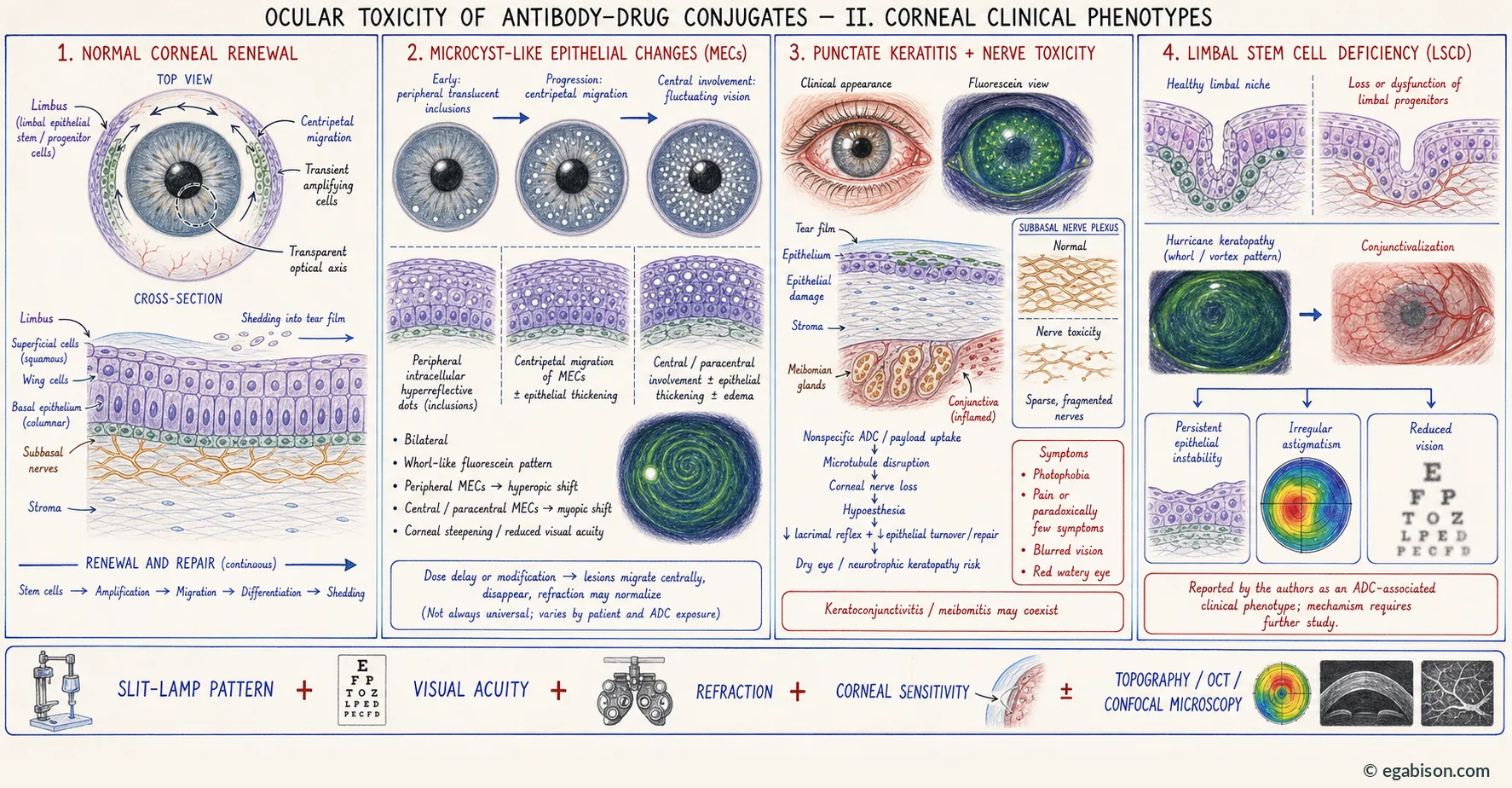
Based on the literature and our clinical experience, we observed three distinct clinical phenotypes:
- superficial microcyst-like epithelial changes (MECs) of the cornea — MEC keratopathy;
- superficial punctate keratitis and keratoconjunctivitis;
- limbal stem cell deficiency (LSCD).
5.1. Microcyst-like epithelial change keratopathy
MECs are bilateral lesions of the corneal epithelium that typically present early after treatment initiation as translucent epithelial inclusions in the corneal periphery on slit-lamp examination, with a whorl-like pattern on fluorescein staining under cobalt blue light.
Although these intraepithelial lesions start in the corneal periphery, they may migrate towards the central cornea and promote epithelial thickening. This centripetal migration is the key element of monitoring: it reflects both the physiological kinetics of epithelial renewal from the limbus and the moment at which the lesion becomes visually significant.
Refractive corollary
A retrospective case series of 29 patients11 with ADC-related keratopathy demonstrated that MECs were significantly associated with hyperopic shifts — peripheral MECs — and myopic shifts — paracentral and central MECs — which corresponded to corneal steepening and resulted in reductions in visual acuity. A recent retrospective study confirmed that vision fluctuations are a major sign of MECs migrating to the centre of the cornea and requiring particular attention.
Refraction is not an ancillary test in this setting: it is an instrument of functional topography. A hyperopic shift signals peripheral involvement; a myopic shift signals paracentral or central migration and should be treated as a warning sign, irrespective of the absolute value of best-corrected visual acuity. Refraction therefore belongs in the monitoring work-up on the same footing as the slit lamp.
5.2. Superficial punctate keratitis and keratoconjunctivitis
Keratitis and keratoconjunctivitis present as red, watery eyes that are sensitive to light with potentially impaired vision, and are caused by underlying ocular surface inflammation.
A case of keratoconjunctivitis combining pseudomembranous conjunctivitis, meibomitis and subsequent diffuse superficial punctate keratitis associated with tisotumab vedotin has been reported. This combination — conjunctival involvement, meibomian gland involvement and keratitis — underscores that toxicity is not confined to the corneal epithelium but engages the ocular surface functional unit as a whole.
5.3. Limbal stem cell deficiency and hurricane keratopathy
LSCD has not yet been reported in the literature, to our knowledge, as a consequence of ADC treatment. However, we had to manage cases of ADC-associated hurricane keratopathy, indicative of LSCD. This type of keratopathy is characterised by a whorl pattern and occurs secondary to a previous disappearance — for example traumatic — of corneal epithelial cells that can be transitory or more persistent in the case of LSCD.
A case of hurricane keratopathy in a female patient in her sixties following treatment with the TROP2-directed datopotamab deruxtecan for lung adenocarcinoma was first reported in this article.
Distinguishing transient hurricane keratopathy from established LSCD is prognostically fundamental. The former reflects centripetal epithelial repopulation after an insult and resolves as renewal is restored. The latter implies damage to the limbal niche itself, with conjunctivalisation, superficial neovascularisation, persistent epithelial instability and irregular astigmatism. The distinction rests on persistence, on the presence of signs of conjunctivalisation and, where necessary, on in vivo confocal microscopy and mapping of epithelial phenotypic expression.
ADC-induced LSCD, should it be confirmed, represents a toxicity of a different order from MEC keratopathy: it does not share the same reversibility. MEC keratopathy generally regresses on treatment withholding or dose reduction; damage to the limbal progenitor niche may leave lasting functional sequelae. This distinction should weigh in the multidisciplinary discussion on continuing treatment.
5.4. Corneal nerve toxicity
Case series studies have documented corneal nerve toxicity, severe reduction of subbasal corneal nerve fibre density and reduced corneal sensitivity with certain ADCs, such as belantamab mafodotin and the investigational drug depatuxizumab mafodotin10. It is important to note that belantamab mafodotin was initially FDA approved for relapsed or refractory multiple myeloma under accelerated approval, but was removed from the US market following failure to meet requirements of the confirmatory phase 3 trial.
Loss of corneal nerves induces corneal hypoaesthesia and thus reduced lacrimal reflex, together with reduced epithelium turnover and repair. Both aspects pave the way for the development of dry eye disease, and eventually, if the process persists, to neurotrophic keratopathy, with potentially severe persistent epithelial defect or even corneal perforation.
The pathophysiology underlying corneal nerve toxicity following belantamab mafodotin treatment, which has an auristatin payload, may be explained by the nonspecific uptake of the ADC in the nerves, resulting in disruption of microtubules and ultimately neurodegeneration. This is supported by in vitro studies showing that MMAE binds30 broadly to microtubules, inhibiting proliferation and mitosis and causing widespread microtubule damage.
Nonspecific ADC uptake in nerve fibres → microtubule disruption → neurodegeneration → corneal hypoaesthesia → reduced lacrimal reflex and epithelial turnover → dry eye → neurotrophic keratopathy → persistent epithelial defect, risk of stromal melt and perforation. This is the only sequence described in this chapter whose endpoint may be a surgical emergency.
Practical consequence: caution with topical non-steroidal anti-inflammatory drugs
In a patient with corneal hypoaesthesia and impaired epithelial renewal, prescribing topical NSAIDs for symptomatic relief carries a risk of stromal melt. We reported a corneal perforation occurring under topical diclofenac in which matrix metalloproteinase induction was localised to the subepithelial zone of the ulcer, rather than diffuse across the whole cornea.[1] This localisation argues against the excipient hypothesis: the French diclofenac formulation involved contains no tocophersolan. The determinant is therefore pharmacological rather than excipient-related, which makes the precaution transposable to topical NSAIDs as a class. Metalloproteinase induction during corneal repair, and the role of EMMPRIN/CD147 in the epithelial-stromal crosstalk that governs it, have been detailed elsewhere.[2]
Figures 3 and 4 correspond to the author's own observations, previously unreported before their publication in Prog Retin Eye Res 2024;103:101302.
Noncorneal adverse events
Albeit less frequent than corneal adverse events, noncorneal adverse events have been reported with ADC use.
As mentioned previously, pseudomembranous conjunctivitis and meibomitis with a good clinical response to topical steroids have been reported in a patient treated by tisotumab vedotin. In addition, cataracts have also been noted with ADC use: cataract was among the grade 3 eye disorder events, as defined using National Cancer Institute CTCAE40 criteria, reported in two patients in a study of 28 patients28 receiving trastuzumab emtansine for advanced HER2+ breast cancer.
The spectrum of noncorneal events reported in trials includes, depending on the agent: conjunctivitis, increased lacrimation, xerophthalmia, photophobia, orbital cyst, retinal haemorrhage, retinal exudates and bacterial conjunctivitis. The wide heterogeneity in reporting detail and the frequent absence of systematic ophthalmic examination make these figures difficult to interpret as true incidences.
Differential diagnosis
Recognising ADC-related toxicity requires excluding competing causes of ocular surface disease in an oncology patient, several of which frequently coexist:
- Other concomitant targeted therapies: consideration should be given to the involvement of any other targeted therapies used in combination with the ADC, given their association with ocular adverse events. MEK, EGFR and FGFR inhibitors and immune checkpoint inhibitors each have their own semiology.
- Conventional chemotherapies: cytarabine (microcystic keratopathy and conjunctivitis), docetaxel (canalicular stenosis and epiphora), 5-fluorouracil.
- Ocular graft-versus-host3 disease in allografted haematology patients, whose severe corneal forms may mimic drug toxicity and whose management differs.[3]
- Pre-existing dry eye disease, common and often aggravated by general condition, age and concomitant treatments.
- Blepharitis and meibomian gland dysfunction, whose worsening under treatment may be mistaken for direct corneal toxicity.
This distinction is not academic: in real-world clinical practice, referral to an ophthalmologist when ocular symptoms develop or worsen may be sufficient to help differentiate ADC-related ocular adverse events from those from other causes.