
Prostaglandins, the COX pathway and 12-HHT/BLT2
This is the question whose answer has changed most since 2003. In short: classical prostanoids are not the driver of corneal repair; the cyclo-oxygenase pathway is — but through products other than prostaglandins. This shift invalidates the canonical explanation of NSAID-related delayed healing and opens an identified therapeutic target.
COX-1 and COX-2 in the cornea
Differential expression of the two isoforms during corneal repair is established. The best-characterised upstream trigger comes from the Bazan group: PAF accumulates in proportion to injury severity and induces COX-2 transcription in the epithelium through a calcium-dependent pathway. COX-2 induction is also documented under UVB stress, in herpetic stromal keratitis and in dry eye disease. One detail is remarkable: COX-2 and PGE synthase are not co-expressed in the same cells. Biosynthesis is therefore transcellular and cooperative.
PGE2 is not the driver of re-epithelialisation
The result is counter-intuitive and rests on direct lipidomic measurement. Liclican et al. compared, in mice, an acute injury (epithelial abrasion) and a chronic injury (sutures)[Liclican 2010]:
- PGE2 does not increase after acute epithelial abrasion. It rises 4.3- to 6.6-fold in chronic injury only (774-1,123 pg/cornea versus 147 pg at baseline).
- Topical PGE2 does not alter healing in the acute phase; in chronic inflammation it amplifies leucocyte infiltration (× 4.5) and neovascularisation (+ 54%).
To which an older and rarely cited result must be added: PGE2 inhibits EGF-induced MAP kinase activity and corneal epithelial proliferation. PGE2 is therefore not a corneal epithelial mitogen; it opposes the canonical mitogenic signal. Recent work adds nuance by showing that a moderate dose of PGE2 promotes repair after alkali burn via annexin A1 induction, suggesting a bell-shaped curve consistent with the acute/chronic dichotomy.
It is commonly stated that NSAIDs delay healing by suppressing PGE2, assumed to be cytoprotective. That reasoning presupposes that PGE2 rises after a wound.
It does not. After acute abrasion its level stays at baseline. There is therefore nothing to suppress, and the explanation collapses.
It is nonetheless still repeated, including in recent reviews.
Mapping the EP receptors
| Layer | Dominant receptors | Coupling |
|---|---|---|
| Corneal epithelium | EP1 (the most strongly expressed in the whole eye), EP2 marked; EP3 and EP4 minimal | Gq / calcium; Gs / cAMP |
| Stroma, keratocytes | EP3 and EP4 predominant | Gi (↓cAMP) / Gs (↑cAMP) |
| Endothelium | EP3 and EP4 strong, FP moderate | — |
Signalling logic is therefore opposite between epithelium and stroma[Schlötzer-Schrehardt 2002]. And tissue sensitivity to PGE2 is largely post-receptor: in chronic injury, EP2 expression rises 14-fold and EP4 8-fold.
The true COX-dependent mediator: 12-HHT and the BLT2 receptor
This work reframes the question[Iwamoto 2017].
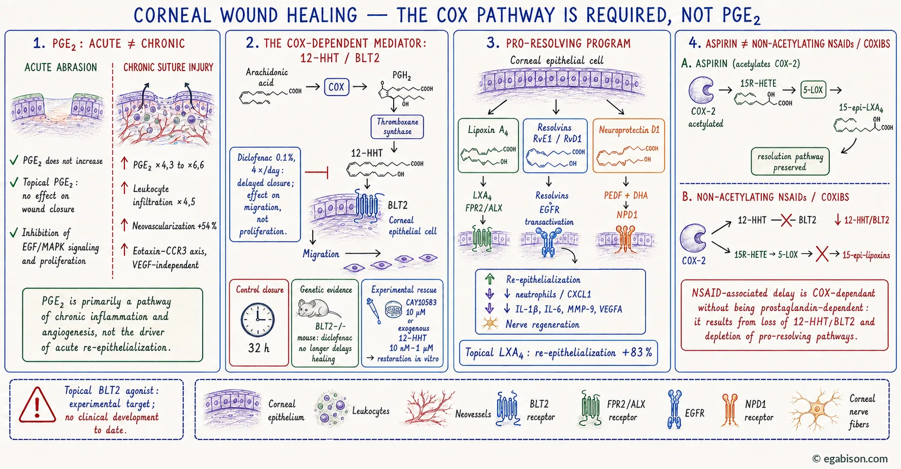
- Model — BALB/c mice, topical diclofenac 0.1% four times daily; HCET lines and primary human corneal epithelial cells in scratch assay.
- Result — complete closure in 32 h in controls, significantly delayed under diclofenac; the effect is on migration, not proliferation.
- Mechanism — NSAIDs suppress COX-dependent production of 12-HHT (12-hydroxyheptadecatrienoic acid), a metabolite derived from PGH2 by thromboxane synthase and the endogenous ligand of the BLT2 receptor. BLT2 is expressed in corneal and conjunctival epithelium.
- Genetic proof — in BLT2−/− mice, diclofenac no longer delays healing.
- Pharmacological rescue — the BLT2 agonist CAY10583 accelerates healing under diclofenac; exogenous 12-HHT restores closure in vitro.
NSAID-related delayed healing is COX-dependent without being prostaglandin-dependent. The target is not a prostaglandin but a non-prostanoid metabolite of the same enzyme. Two corollaries follow. First, the mechanism is vehicle-independent, which fits the refutation of the excipient hypothesis. Second, an identified therapeutic target exists: a topical BLT2 agonist, or 12-HHT itself, would restore healing without abolishing the desired anti-inflammatory effect. No clinical development has been undertaken to date.
The lipid signals of repair: lipoxins, resolvins, neuroprotectin D1
These three families derive from the same precursors as prostaglandins, but through distinct enzymatic branches. They are produced by the injured epithelium itself and act through their own receptors — FPR2/ALX for lipoxins, EGFR transactivation for resolvins. This is the best-supported area in all of corneal lipid biology, driven by the Serhan, Gronert and Bazan groups[Gronert 2005].
Lipoxin A4
An intrinsic mediator of the ocular surface, whose receptor FPR2/ALX is expressed by many healthy corneal and conjunctival cell types; corneal epithelial cells are the principal source of endogenous pro-resolving mediators. Topical LXA4 increases re-epithelialisation by 83%, reduces CXCL1 and neutrophil infiltration, and in severe alkali burn reduces opacity, neovascularisation, IL-1β, IL-6, MMP-9 and VEGFA. Notably, epithelial removal abolishes corneal Alox15 expression, restored during recovery — injury transiently suppresses the tissue's pro-resolving capacity.
Resolvins and docosanoids
RvE1 accelerates wound closure in human corneal epithelium comparably to EGF; RvD1 controls dendritic maturation and suppresses alloimmunity in corneal transplantation. Importantly, resolvin-induced epithelial migration depends on EGFR transactivation — the mechanism habitually attributed to PGE2 is, in this tissue, that of pro-resolving mediators.
The combination PEDF + DHA increases corneal nerve area 2.5-fold, at 2 and 4 weeks after stromal dissection. Neuroprotectin D1 levels are quadrupled. PEDF alone and DHA alone have no effect. In diabetic mice, PEDF + DHA increase nerve regeneration, sensitivity and tear production.
Aspirin, non-acetylating NSAIDs and coxibs
COX-2 acetylated by aspirin no longer produces prostaglandins but generates 15R-HETE, converted by 5-LOX into 15-epi-lipoxins, which are more resistant to degradation — "lipid mediator class switching". Non-acetylating NSAIDs and coxibs merely inhibit COX-2: they therefore suppress both 12-HHT/BLT2 and generation of 15-epi-lipoxins. A coxib may be doubly deleterious for the ocular surface where aspirin preserves a resolution pathway.
Translational relevance: 15-epi-LXA4 is present in human emotional tears, and human corneas stored overnight in 100 nM 15-epi-LXA4 before Optisol-GS show graft viability rising from 36 to 56%.
What is left of the lipoxygenase "shunt"?
The mechanism is classical: COX inhibition redirects arachidonic acid towards 5-lipoxygenase, producing leukotrienes and HPETEs, neutrophil chemoattractants, hence delivery of MMP-8, MMP-9 and elastase. Two serious objections must nonetheless be raised:
- If the pro-inflammatory shunt were the principal driver of delayed healing, BLT2 knockout should not have abolished it. Yet it abolishes it completely[Iwamoto 2017]. The shunt cannot therefore be the sole explanation.
- The 12/15-LOX pathway has a beneficial arm: it promotes epithelial healing and host defence. Describing the "shunt to lipoxygenase" as a uniformly deleterious mechanism is a simplification.
Prostaglandins in the strict sense are not important for acute corneal healing — they do not rise after abrasion, they inhibit EGF-induced epithelial proliferation, and their topical application does not accelerate closure. They are the circuitry of chronic inflammation, pathological angiogenesis and probably pain.
The cyclo-oxygenase pathway is important — but through a non-prostanoid metabolite, 12-HHT, a BLT2 agonist, whose loss suffices to explain delayed epithelial migration and whose restoration abolishes it.
The common statement that "NSAIDs prevent healing because they block prostaglandins" is therefore imprecise: the delay is due to cyclo-oxygenase inhibition, not to loss of prostaglandins.