
Ranked mechanisms and at-risk patients
Whatever the initial cause, sterile keratolysis follows a single circuit. This is not passive digestion but an imbalance between matrix deposition and degradation, governed by the MMP/TIMP ratio and controlled at the epithelial-stromal interface.
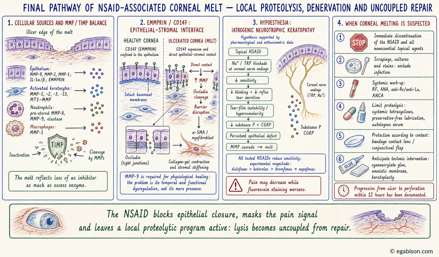
Cellular sources
- Epithelium — chiefly MMP-9, plus MMP-2 and MMP-1; releases IL-1α/β and EMMPRIN, which activate the stroma. Dissolution of the basement membrane precedes and conditions stromal lysis.
- Activated keratocytes — MMP-1 (the only collagenase able to attack native fibrillar collagen I/III), MMP-2, MMP-3 (stromelysin-1, an upstream activator), MMP-13, MT1-MMP.
- Neutrophils — MMP-8 and MMP-9 pre-stored in granules, hence releasable without transcription, which explains the abruptness of neutrophil-driven melts; plus elastase, which degrades proteoglycans and inactivates TIMPs, releasing the brake.
- Macrophages — a source of MMP-1 in rheumatoid keratolysis.
The clearest demonstration remains that of Riley et al. on 8 ulcerated corneas from rheumatoid arthritis[Riley 1995]. MMP-1 is expressed by stromal cells in 8 corneas out of 8, and absent from normal tissue. TIMP-1 does the opposite: abundant in normal stroma, greatly reduced or absent in diseased corneas. Melt is as much a loss of inhibitor as an excess of enzyme.
EMMPRIN/CD147, the control point of the epithelial-stromal interaction
CD147 is normally confined to the corneal epithelium; in ulcerated corneas its expression is altered and extended, and direct epithelium-keratocyte contact induces MMPs[Gabison 2005][Gabison 2009]. The mechanism goes beyond proteolytic induction. EMMPRIN promotes myofibroblast differentiation, by inducing α-SMA and collagen gel contraction[Huet 2008]. It also modulates epithelial barrier function, through MMP-dependent cleavage of occludin[Huet 2011]. This is the link connecting proteolysis to tight junction disruption.
In an avian model, corticosteroids and NSAIDs worsened the lesion within 5 days. MMP-9 expression, increased 2.5-fold in the normally healing group, became undetectable under both classes. MMP-9 is therefore necessary for physiological healing.
Melt is not simple "MMP overexpression". It is the temporal and functional dysregulation of a programme that stays spatially in its proper place: induced at the healing front and in the injured anterior stroma, with a central-to-peripheral gradient. In other words, exactly where repair should occur — and does not.
The distinction has practical consequences: it directs the target towards restoring epithelial closure and controlling the MMP/TIMP ratio at the injured site, rather than towards eliminating a hypothetical surface toxin.
Induced corneal hypoaesthesia: an iatrogenic neurotrophic keratopathy
This hypothesis, examined and then set aside in 2001, is reopened by data from 2012-2017.
The pharmacological basis
Independently of COX inhibition, NSAIDs directly modulate voltage-gated Na⁺, Ca²⁺ and K⁺ channels, TRP channels and chloride channels; authors propose diclofenac as a template channel modulator. NSAIDs block compound action potentials in isolated nerve — that is, they possess intrinsic local anaesthetic activity.
Human aesthesiometry data
| Study | Population | Result |
|---|---|---|
| Sun & Gimbel 1997 | Normal corneas | Measurable reduction under ketorolac and diclofenac. |
| Aragona 2000[Aragona 2000] | 90 healthy subjects | Only diclofenac induced a significant reduction persisting at 1 week (p<0.001), with no visible epithelial lesion. |
| Aragona 2005[Aragona 2005] | 20 randomised Sjögren's patients with keratitis | Significant reduction in sensitivity at day 30 (p<0.05), more marked under diclofenac. Reduced discomfort from day 15. But fluorescein staining significantly worsened under diclofenac 7 days after stopping (p=0.02). |
| Singer 2015[Singer 2015] | 10 healthy volunteers | All NSAIDs significantly reduced sensitivity (p<0.001). Magnitude: diclofenac 28.6 mm > ketorolac 21.1 mm > bromfenac 16.9 mm > nepafenac 16.4 mm. |
The causal chain
Topical NSAID → Na⁺/TRP blockade of corneal nerve endings → ↓ sensitivity → ↓ blink rate and ↓ reflex tear secretion → tear film instability, hyperosmolarity, exposure keratopathy → ↓ neuronal release of substance P and CGRP, epithelial trophic factors → persistent epithelial defect → MMP cascade → melt.
Guidera, Luchs and Udell explicitly examined and then set aside the neurotrophic analogy with anaesthetic abuse, on the grounds that NSAID analgesia was then considered purely anti-inflammatory. The 2012-2017 ion channel data remove that argument. The analogy is all the more legitimate in that anaesthetic abuse combines exactly three components — suppression of the protective reflex, direct epithelial toxicity, repeated instillation with preservative — which topical NSAIDs also combine, at lower intensity. And histology of the Bordeaux case showed total destruction of corneal innervation[Mortemousque 2002].
Direct cellular toxicity
ROS/p53-dependent apoptosis of human corneal epithelial cells under diclofenac, independent of COX; direct inhibition of rabbit epithelial migration, and not of proliferation — consistent with the 12-HHT/BLT2 mechanism discovered sixteen years later. To which is added uncoupling of oxidative phosphorylation: NSAIDs are lipophilic weak acids behaving as protonophores, dissipating the mitochondrial proton gradient. This is the canonical explanation of NSAID enteropathy; transposed to an avascular epithelium with strictly aerobic metabolism, the hypothesis is plausible but has never been explored and must be presented as extrapolation.
Clinical masking and synergy with corticosteroids
Masking is clinically demonstrated by the dissociation in Aragona 2005: discomfort falls significantly by day 15 while fluorescein staining worsens and persists after stopping. Clinically it produces misleading pictures: Asai describes melts of 60 to 80% depth with "discrete" hyperaemia and infiltration[Asai 2006].
Combination with corticosteroids is the most consistent iatrogenic risk factor (≥10/16 of Guidera's patients). The two effects add up through distinct mechanisms. The corticosteroid inhibits keratocyte proliferation and migration and re-epithelialisation: it prevents repair. The NSAID blocks epithelial migration via 12-HHT/BLT2, dysregulates MMPs and removes the alarm signal: it accelerates destruction and masks it. Uncoupling of lysis from repair becomes complete.
Ranking of mechanisms by level of evidence
| Level | Mechanism | Pivotal reference |
|---|---|---|
| High | Collapse of 12-HHT → loss of BLT2 signalling → inhibition of epithelial migration (genetic knockout + pharmacological rescue) | Iwamoto 2017 |
| High | Induced corneal hypoaesthesia, with symptom/lesion dissociation (randomised trials) | Aragona 2000, 2005; Singer 2015 |
| High | Local, dysregulated MMP induction at the healing front and in the injured anterior stroma | Gabison 2003; O'Brien 2001 |
| Moderate | ROS/p53-dependent epithelial apoptosis (in vitro) | — |
| Moderate | Direct blockade of Na⁺, K⁺, Ca²⁺ and TRP channels — intrinsic local anaesthesia | Gwanyanya 2012; Tsagareli 2017 |
| Moderate | Lipoxygenase shunt → LTB4/HPETE → neutrophil chemotaxis (contradicted as the principal mechanism by BLT2 knockout) | Ku 1986 |
| Low | Loss of cytoprotective PGE2 — the canonical mechanism, not supported by direct lipidomic measurement in acute injury | Liclican 2010 (against) |
| Low | Mitochondrial uncoupling (extrapolation from non-corneal tissue) | Mahmud 1996 |
| Refuted | Toxicity of the excipient tocophersolan — never demonstrated in cornea; refuted by cases occurring without it | Chapter 2 |
At-risk patients
Dry eye and Sjögren's syndrome
The substrate of fragility is explicitly the same as the final pathway of melt. The TFOS DEWS II report describes the cascade[TFOS DEWS II 2017]. Tear hyperosmolarity reaches 800 to 1,900 mOsm/L in hot spots, at film break-up sites. It triggers transcriptional activation of inflammatory MMPs, chiefly MMP-9, via MAPK and NF-κB. MMP-9 then cleaves occludin and disrupts the epithelial barrier.
The NSAID adds to this substrate blockade of epithelial migration, MMP dysregulation and — specifically demonstrated in these patients — reduced sensitivity with worsening fluorescein staining persisting after withdrawal[Aragona 2005]. This is the only randomised prospective evidence available in dry eye, and it is unfavourable. In a series of 46 eyes with corneal ulceration in Sjögren's, ulceration was the first revealing sign of the disease in 26% of patients.
Ocular graft-versus-host disease (GVHD)
This is currently the only cohort documenting NSAID imputability in these patients, and it changes the order of magnitude of baseline risk[Bourdin 2025]. Among 140 patients at a tertiary centre:
— 33 patients (23.6%) developed corneal ulceration or perforation. That is nearly five times the usually cited proportion. Median latency was 39 months after transplantation.
— Overall survival did not differ between severe and non-severe ocular involvement. Severe corneal involvement is therefore not merely a marker of systemic severity: it is an autonomous ophthalmological problem.
— And above all: "a high proportion of complications occurred after non-steroidal anti-inflammatory drug (NSAID) treatments".
The authors' conclusion is the most categorical in the entire literature: patients with GVHD "should not, in any case, be treated with local ocular NSAIDs, due to the severity of potential complications".
Benefit/risk. Ocular GVHD is the only setting with data on both sides. On the benefit side: none — "although topical non-steroidal anti-inflammatory drugs are used in oGVHD, there is no evidence for their efficacy". On the risk side: 23.6% ulceration or perforation. This is the only situation in which the literature justifies proscription rather than a simple warning — a position no SmPC or learned society has yet adopted.
Neurotrophic keratopathy
Denervation abolishes release of substance P, CGRP and NGF, and impairs epithelial renewal. Relevant aetiologies are herpes and shingles, diabetes, refractive and corneal surgery, and — explicitly — "chronic use of topical medications". Saad et al.'s series (Fondation Rothschild) covers 63 eyes: overall healing rate 79.5%, mean latency 44.8 days[Saad 2020]. In other words, one eye in five fails to heal. And exposure to a harmful topical agent is measured in weeks. The authors' position is explicit: all topical medications should be stopped, and NSAIDs "should be avoided".
The aggravating loop is specific: NSAIDs are themselves corneal anaesthetics. On an already hypoaesthetic cornea, the NSAID worsens functional denervation and removes the warning signal — which explains pauci-symptomatic presentations.
Diabetes
Diabetic keratopathy combines sensory neuropathy, chronic inflammation, abnormal basement membrane adhesion and AGE accumulation, resulting in impaired re-epithelialisation. A validated risk factor: 4/16 of Guidera's patients, first-ranked trigger in Rigas, and explicitly listed in European SmPCs and the Medsafe alert.
The most demonstrative case combines five factors. A 50-year-old woman, keratoconus, pachymetry 374-408 µm, undergoing crosslinking. Nepafenac prescribed for one week, continued for six without review. HbA1c 9.1%. The result: a 2 × 2 mm perforation with iris herniation at six weeks, requiring emergency tectonic keratoplasty[Mohamed-Noriega 2016].
Systemic autoimmunity
Peripheral ulcerative keratitis is classically associated with rheumatoid arthritis — the commonest systemic cause —, granulomatosis with polyangiitis, relapsing polychondritis, lupus and vasculitides. The mechanism is identical to the final MMP-dependent pathway of NSAID toxicity, giving pharmacological additivity rather than mere coincidence. As with Sjögren's, the underlying disease is sometimes revealed by the accident.
Cases without identifiable risk factors
They exist, but they are few. And none involves a strictly intact cornea without an iatrogenic cofactor. The two cleanest demonstrations are these. Feiz 2009: a 35-year-old man, uncomplicated bilateral PRK, no history — but a PRK epithelial defect and intensive dosing[Feiz 2009]. Mikropoulos 2024: a 70-year-old woman "previously healthy", perforation after 2 days of an NSAID combined with an antiseptic, in preparation for cataract surgery[Mikropoulos 2024].
In a meta-analysis of 38 published cases, 16 rows carried the mention "none reported" — 42% —, the authors retaining about 20% after weighting and noting that the mention may reflect incomplete documentation. Three observations demonstrate that "no known risk factor" does not equal "no risk factor": two cases of Sjögren's and one of rheumatoid arthritis diagnosed after the perforation.