
Recruitment & polarization signals
The recruitment of circulating monocytes and their polarization into functional macrophages are orchestrated by chemical and mechanical signals specific to the injured corneal microenvironment [Yaghmour 2026].
Epithelial injury releases DAMPs (HMGB1, ATP) and pro-inflammatory cytokines, generating a chemokine gradient — CCL2/MCP-1, CCL5/RANTES, CX3CL1 (fractalkine) — relayed by M-CSF and IL-1β/TNF-α. Circulating monocytes adhere to the limbal vascular endothelium and then transmigrate to the injury site; local hypoxia further amplifies this recruitment.
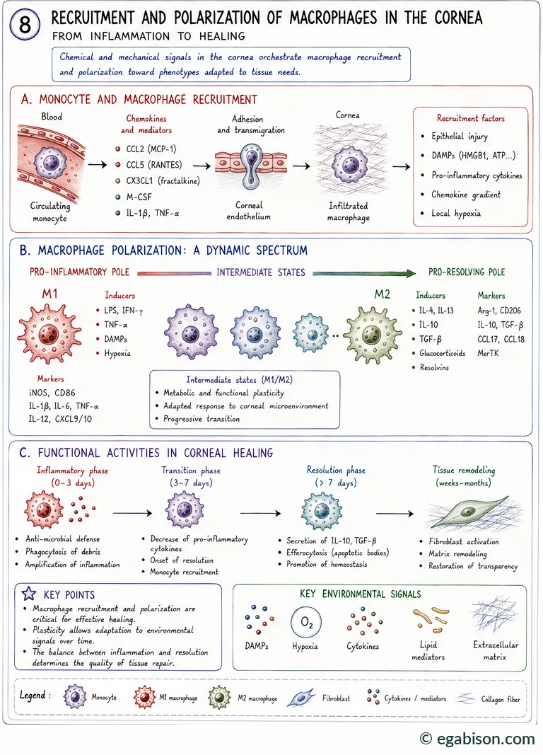
Once recruited, macrophages polarize along a functional spectrum, historically simplified into two poles:
Between these two poles, intermediate states reflect metabolic and functional plasticity: the response adapts continuously to the corneal microenvironment rather than flipping between fixed states. It is precisely this plasticity that the M1/M2 classification, useful as a conceptual starting point, does not fully capture [Martinez & Gordon 2014].
The M1/M2 framework remains pedagogically useful, but the review by Yaghmour et al. stresses that it is a simplification of a much richer continuum of macrophage programmes, shaped by tissue context, signal kinetics and local cellular interactions.
Transcriptomic states: a continuum
Single-cell transcriptomic profiling has revealed substantial heterogeneity among corneal macrophage populations, organized not into two sharp categories but into a five-state continuum: pro-inflammatory M1 → intermediate state 1 → intermediate state 2 → pro-resolving M2 → pro-fibrotic M2 [Yaghmour 2026].
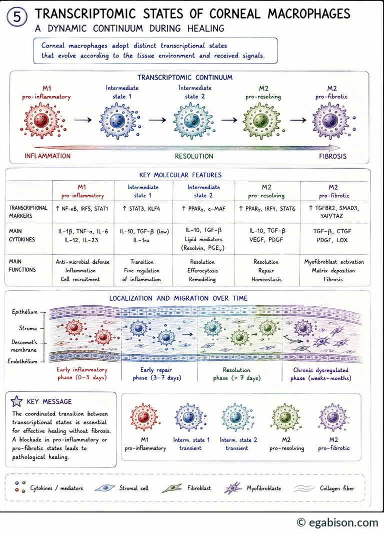
| State | Transcriptional markers | Main cytokines | Main functions |
|---|---|---|---|
| Pro-inflammatory M1 | NF-κB, IRF5, STAT1 ↑ | IL-1β, TNF-α, IL-6, IL-12, IL-23 | Antimicrobial defence, inflammation, cell recruitment |
| Intermediate 1 | STAT3, KLF4 ↑ | IL-10, TGF-β (low), IL-1ra | Transition, fine regulation of inflammation |
| Intermediate 2 | PPARγ, c-MAF ↑ | IL-10, TGF-β, lipid mediators (resolvins, PGE₂) | Resolution, efferocytosis, remodeling |
| Pro-resolving M2 | PPARγ, IRF4, STAT6 ↑ | IL-10, TGF-β, VEGF, PDGF | Resolution, repair, homeostasis |
| Pro-fibrotic M2 | TGFBR2, SMAD3, YAP/TAZ ↑ | TGF-β, CTGF, PDGF, LOX | Myofibroblast activation, matrix deposition, fibrosis |
These states are also distributed in space and time: pro-inflammatory macrophages predominate in the epithelium and superficial stroma during the early phase (0-3 days); intermediate states progressively migrate deeper during early repair (3-7 days); pro-resolving macrophages settle in at resolution (> 7 days); if this transition fails, pro-fibrotic macrophages persist adjacent to Descemet's membrane and the endothelium, weeks to months later.
The coordinated transition between these transcriptional states is essential for effective, non-fibrotic healing. A blockade in either the pro-inflammatory or the pro-fibrotic state leads, in both cases, to pathological healing.
M2 pro-resolving subtypes
M2 macrophages are not a single entity: they subdivide into specialized subpopulations with complementary functions, defined by their inducing signals [Yaghmour 2026].
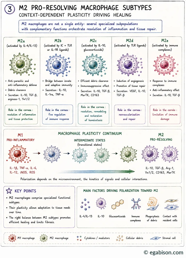
M2a to M2eff: five profiles
| Subtype | Activating stimulus | Primary function |
|---|---|---|
| M2a (classical) | IL-4, IL-13 | Wound healing, collagen synthesis; prolonged activity may promote fibrosis |
| M2b (immune regulator) | Immune complexes + TLR | Mixed pro- and anti-inflammatory response; regulates the magnitude of the immune response rather than suppressing it |
| M2c (deactivation) | IL-10, TGF-β, glucocorticoids | Matrix remodeling, immunosuppression, efficient efferocytosis — terminates inflammation |
| M2d (angiogenic) | TLR ligands, A2a agonists | Promotes angiogenesis; double-edged role, pathological if regulatory constraints fail |
| M2eff (efferocytic) | Phagocytosis of apoptotic cells | Clearing cellular debris, secretion of IL-10/TGF-β post-inflammation |
The M2d subtype illustrates the ambivalence of M2 macrophages well: its pro-angiogenic capacity can favour tissue repair in some contexts, but it is also the mechanism by which macrophages breach the cornea's avascular privilege when regulatory signals fail — opening the door to pathological neovascularization.
The right balance between these subtypes, more than the isolated presence of any one of them, determines effective repair: resolution of inflammation, orderly matrix remodeling and restoration of corneal transparency.
Glossary of abbreviations used in this course
Scientific abbreviations and acronyms used throughout the 10 pages of this course, listed alphabetically.
No matching term.
- 95% CI
- 95% confidence interval
- AAV
- adeno-associated virus (gene-therapy viral vector)
- ABCG2
- ABCG2 transporter, a limbal stem-cell marker
- AMT
- amniotic membrane transplantation
- anti-VEGF
- anti-angiogenic treatment targeting VEGF
- ASC
- inflammasome adaptor protein (Apoptosis-associated Speck-like protein containing a CARD)
- BrdU
- bromodeoxyuridine, a cell-proliferation marker
- CCL2
- CCL2 chemokine, synonym of MCP-1; recruits circulating monocytes
- CCR2
- receptor for CCL2; marker of newly recruited monocytes/macrophages
- CD147
- cluster of differentiation 147; synonym of EMMPRIN and basigin
- CD163
- M2 macrophage marker; scavenger receptor for haemoglobin-haptoglobin
- CD206
- mannose receptor; marker of M2 polarization
- CD74
- cell-surface receptor for MIF
- CDVA
- corrected distance visual acuity
- CGRP
- calcitonin gene-related peptide
- CTGF
- connective tissue growth factor
- CX3CL1
- fractalkine, a chemokine involved in monocyte/macrophage recruitment
- CXCR2/CXCR4
- chemokine receptors, also receptors for MIF
- DAMP
- damage-associated molecular pattern
- DESI
- direct epithelial–stromal interaction
- EGF
- epidermal growth factor
- EGFR
- epidermal growth factor receptor
- EMMPRIN
- extracellular matrix metalloproteinase inducer; synonym of CD147
- ETDRS
- reference visual-acuity scale used in clinical research (Early Treatment Diabetic Retinopathy Study)
- FasL
- Fas ligand, an apoptosis-inducing protein
- FISH
- fluorescence in situ hybridisation
- GAG
- glycosaminoglycan(s)
- GM-CSF
- granulocyte-macrophage colony-stimulating factor
- GVHD
- graft-versus-host disease
- HGF
- hepatocyte growth factor
- HIF-3α
- hypoxia-inducible factor, 3-alpha subunit
- HMGB1
- high mobility group box 1, an alarmin released by injured cells (DAMP)
- IFN-γ
- interferon gamma
- IGF-1
- insulin-like growth factor 1
- IL-1
- interleukin-1
- iNOS
- inducible nitric oxide synthase
- IPAS
- inhibitory PAS domain protein
- KGF
- keratinocyte growth factor
- LASIK
- laser-assisted in situ keratomileusis
- LogMAR
- logarithmic visual-acuity scale
- LOX
- lysyl oxidase, a collagen cross-linking enzyme
- LPS
- bacterial lipopolysaccharide
- M1
- classically activated macrophage, pro-inflammatory profile
- M2
- alternatively activated macrophage, pro-resolving/reparative profile (subtypes M2a-M2d, M2eff)
- MEN2B
- multiple endocrine neoplasia type 2B
- MerTK
- receptor tyrosine kinase involved in efferocytosis
- MIF
- macrophage migration inhibitory factor
- MRI
- magnetic resonance imaging
- NGF
- nerve growth factor
- NK
- neurotrophic keratitis
- NLRP3
- NLRP3 inflammasome (NOD-, LRP- and pyrin domain-containing protein 3)
- NO
- nitric oxide
- NSAID
- nonsteroidal anti-inflammatory drug
- PACAP
- pituitary adenylate cyclase-activating polypeptide
- PAF
- platelet-activating factor
- PAMP
- pathogen-associated molecular pattern
- PDGF
- platelet-derived growth factor
- PED
- persistent epithelial defect
- PEDF
- pigment epithelium-derived factor
- PGE₂
- prostaglandin E2
- PRK
- photorefractive keratectomy (laser surface ablation)
- pro-NGF
- precursor form of NGF
- RGTA
- matrix-therapy ReGeneraTing Agent, e.g. Cacicol
- rhNGF
- recombinant human NGF (cenegermin)
- ROS
- reactive oxygen species
- Smad
- signalling proteins of the TGF-β pathway
- SPK
- superficial punctate keratitis
- sVEGFR-1
- soluble form of VEGF receptor-1 (= sFlt-1)
- sVEGFR-3
- soluble form of VEGF receptor-3
- TGF-β
- transforming growth factor beta
- TIMP
- tissue inhibitor of metalloproteinases
- TLR
- Toll-like receptor
- TNF-α
- tumour necrosis factor alpha
- TRPV4
- transient receptor potential vanilloid 4, a mechanosensitive ion channel
- uPA
- urokinase-type plasminogen activator
- V1
- ophthalmic branch of the trigeminal nerve
- VEGF
- vascular endothelial growth factor
- VEGF-C
- lymphangiogenic isoform of VEGF
- VIP
- vasoactive intestinal peptide
- YAP/TAZ
- Hippo pathway effectors, mechanosensors of matrix stiffness
- α-SMA
- alpha-smooth muscle actin, a myofibroblast marker