
Molecular regulators: NLRP3, MIF, TGF-β
Three major molecular axes orchestrate the macrophage-fibroblast dialogue and determine the outcome of corneal healing [Yaghmour 2026].
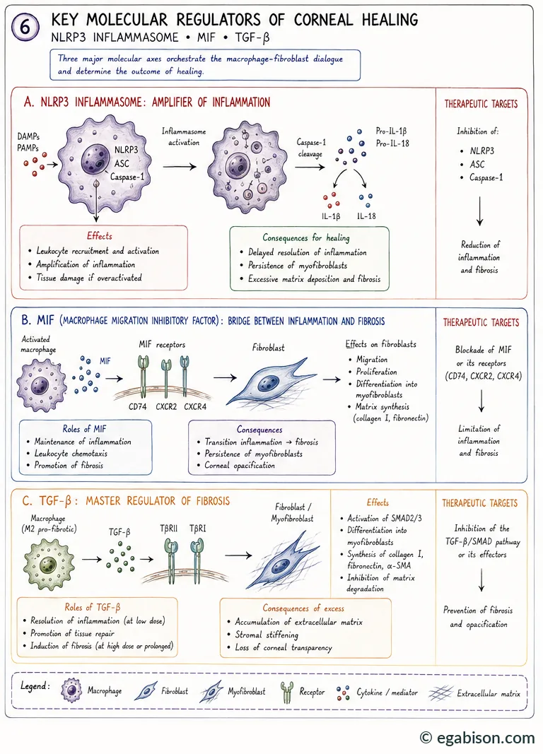
NLRP3: amplifier of inflammation
Activation of the NLRP3 inflammasome by DAMPs and PAMPs triggers assembly of NLRP3-ASC-caspase-1, which cleaves pro-IL-1β and pro-IL-18 into their active forms. In mice, Xu et al. showed that NLRP3 activation in infiltrating macrophages amplifies TGF-β1 expression by the corneal epithelium, creating a feed-forward loop that sustains myofibroblast survival and collagen deposition; genetic or pharmacological inhibition of this pathway (e.g., MCC950) reduces stromal haze in murine models [Xu 2022].
MIF: the pro-fibrotic lock
Macrophage migration inhibitory factor (MIF) acts as an upstream regulator of sustained macrophage activation in ocular tissues. It promotes macrophage retention at the injury site and amplifies pro-inflammatory signalling via the NF-κB and STAT3 pathways, reinforcing IL-1β and TNF-α production while actively opposing deactivation and resolution programmes. MIF thus acts as a true molecular checkpoint that locks macrophages into a pathogenic state — a promising therapeutic target for restoring proper immune timing [Yaghmour 2026]. Its receptors — CD74, CXCR2, CXCR4 — are detectable in ocular fluids in inflammatory contexts [Taguchi 2001].
TGF-β: master cytokine
TGF-β acts through two complementary pathways once bound to its TβRI/TβRII receptors: the canonical Smad2/3 pathway, which activates transcription of pro-fibrotic genes (COL1A1, FN1, α-SMA); and non-canonical pathways (MAPK, Rho/ROCK, PI3K/Akt) that directly drive myofibroblast activation, contraction and matrix deposition. As detailed in the physiology chapter of this course, TGF-β has a double face: essential for resolution of inflammation and repair at low dose, it becomes one of the main drivers of fibrosis when its activity is excessive or prolonged.
NLRP3, MIF and TGF-β converge on the same pathological lock: macrophage inflammation that fails to switch off. Inhibiting them only makes sense within that window of persistence — not during the early inflammatory phase, which is essential for host defence.
Fibrosis & macrophage-fibroblast dialogue
Excessive matrix deposition driven by persistent pro-fibrotic macrophages does more than fill the stroma: it alters its mechanical properties, closing the loop back onto the macrophages themselves.
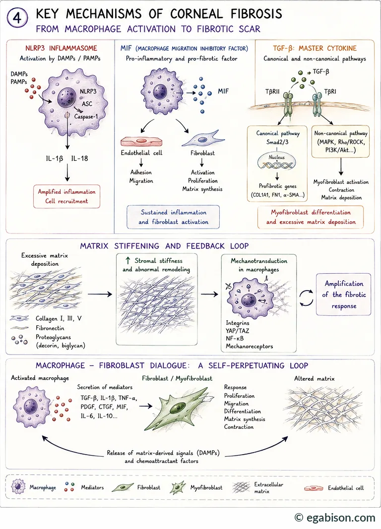
Excess deposition of collagen I, III and V, fibronectin and proteoglycans (decorin, biglycan) stiffens the stroma and disrupts its remodelling. This increased stiffness is sensed by macrophages via integrins, the YAP/TAZ pathway, NF-κB and mechanoreceptors — a form of mechanotransduction that further activates macrophages and amplifies the fibrotic response, independently of any new classical inflammatory signal.
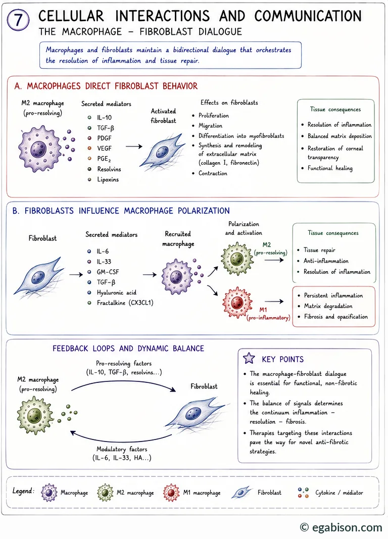
This dialogue is bidirectional. On one side, pro-resolving M2 macrophages secrete IL-10, TGF-β, PDGF, VEGF, PGE₂, resolvins and lipoxins, which direct fibroblasts toward proliferation, migration, controlled differentiation and balanced matrix synthesis — favouring restoration of transparency. On the other, fibroblasts themselves secrete IL-6, IL-33, GM-CSF, TGF-β, hyaluronic acid and fractalkine (CX3CL1), which direct the polarization of newly recruited macrophages — toward a reparative M2 profile in a favourable context, or toward a deleterious M1 profile if the balance tips, with resulting persistent inflammation, matrix degradation and, eventually, fibrosis and opacification.
The macrophage-fibroblast dialogue is essential for functional, non-fibrotic healing. It is the balance of signals — not the existence of this dialogue itself — that determines the inflammation-resolution-fibrosis continuum. Therapies targeting these interactions pave the way for novel anti-fibrotic strategies.
Therapeutic implications, summary & references
Recognizing macrophages as temporal regulators of corneal healing opens the way to a timing-based immunotherapy, rather than global immune suppression [Yaghmour 2026]. Two broad strategies are being explored:
- Macrophage cell therapy (introduction of exogenous or reprogrammed macrophages): an interesting proof of mechanistic concept, but its translational applicability remains uncertain.
- Manipulation of host macrophages — the favoured approach: reprogramming resident and recruited populations in situ, without introducing exogenous cells, through modulation of IL-1/TGF-β/MIF signalling, delivery of immunoregulatory factors, metabolic reprogramming, or mesenchymal stromal cell-derived extracellular vesicles (MSC-EVs). The goal is not to suppress macrophage activity, but to promote a timely transition toward resolution-phase programmes.
| Pathway | Targets / agents | Intended effect |
|---|---|---|
| NLRP3 inflammasome | NLRP3, ASC, caspase-1 (e.g., MCC950) | ↓ Inflammation, ↓ TGF-β, ↓ fibrosis |
| MIF | MIF or its receptors (CD74, CXCR2, CXCR4) | Limiting inflammation and fibrosis |
| TGF-β / Smad | TGF-β/Smad pathway or its effectors | Prevention of fibrosis and opacification |
Summary & key points
- The cornea harbours resident macrophages (surveillance, homeostasis) and recruits monocyte-derived macrophages (acute defence) — two non-redundant roles.
- The response follows a four-phase timeline: inflammation (0-3 days), repair (3-7 days), resolution (> 7 days), or chronic drift (weeks-months) if the transition fails.
- The M1/M2 framework is a simplification of a five-state transcriptomic continuum and of five M2 subtypes (M2a-M2d, M2eff) with distinct functions.
- NLRP3, MIF and TGF-β form a molecular triangle that locks macrophages into a pro-fibrotic state when their activity persists.
- The bidirectional macrophage-fibroblast dialogue, amplified by matrix stiffening (YAP/TAZ mechanotransduction), self-perpetuates fibrosis once triggered.
- The therapeutic target is not macrophage suppression, but restoring their proper timing — favouring a rapid transition toward resolution states.
A macrophage is neither "good" nor "bad" in itself: it is its timing that decides the outcome. A necessary defender on day 1, it becomes the chief architect of fibrosis if it fails — or is unable — to stand down in time.
Original teaching course. Primary source: systematic review by Yaghmour, Arabpour, Al-Khudari & Djalilian (Life, 2026), Department of Ophthalmology and Visual Science, University of Illinois Chicago — open-access article.
Primary source
- Yaghmour A, Arabpour Z, Al-Khudari H, Djalilian A. When Macrophages Heal and When They Scar: Timing in Corneal Fibrosis. Life 2026;16(7):1090. doi:10.3390/life16071090 (open access).
Supporting references cited in this course
- Streilein JW. Ocular immune privilege: therapeutic opportunities from an experiment of nature. Nat Rev Immunol 2003;3(11):879-889.
- Wynn TA, Vannella KM. Macrophages in Tissue Repair, Regeneration, and Fibrosis. Immunity 2016;44(3):450-462.
- Martinez FO, Gordon S. The M1 and M2 paradigm of macrophage activation: time for reassessment. F1000Prime Rep 2014;6:13.
- Mantovani A, Biswas SK, Galdiero MR, Sica A, Locati M. Macrophage plasticity and polarization in tissue repair and remodelling. J Pathol 2013;229(2):176-185.
- Xu J, Chen P, Luan X, et al. The NLRP3 Activation in Infiltrating Macrophages Contributes to Corneal Fibrosis by Inducing TGF-β1 Expression in the Corneal Epithelium. Invest Ophthalmol Vis Sci 2022;63(9):15.
- Taguchi C, Sugita S, Tagawa Y, Nishihira J, Mochizuki M. Macrophage migration inhibitory factor in ocular fluids of patients with uveitis. Br J Ophthalmol 2001;85(11):1367-1371.
- Ambati BK, Nozaki M, Singh N, et al. Corneal avascularity is due to soluble VEGF receptor-1. Nature 2006;443(7114):993-997.
Glossary of abbreviations used in this course
Scientific abbreviations and acronyms used throughout the 10 pages of this course, listed alphabetically.
No matching term.
- 95% CI
- 95% confidence interval
- AAV
- adeno-associated virus (gene-therapy viral vector)
- ABCG2
- ABCG2 transporter, a limbal stem-cell marker
- AMT
- amniotic membrane transplantation
- anti-VEGF
- anti-angiogenic treatment targeting VEGF
- ASC
- inflammasome adaptor protein (Apoptosis-associated Speck-like protein containing a CARD)
- BrdU
- bromodeoxyuridine, a cell-proliferation marker
- CCL2
- CCL2 chemokine, synonym of MCP-1; recruits circulating monocytes
- CCR2
- receptor for CCL2; marker of newly recruited monocytes/macrophages
- CD147
- cluster of differentiation 147; synonym of EMMPRIN and basigin
- CD163
- M2 macrophage marker; scavenger receptor for haemoglobin-haptoglobin
- CD206
- mannose receptor; marker of M2 polarization
- CD74
- cell-surface receptor for MIF
- CDVA
- corrected distance visual acuity
- CGRP
- calcitonin gene-related peptide
- CTGF
- connective tissue growth factor
- CX3CL1
- fractalkine, a chemokine involved in monocyte/macrophage recruitment
- CXCR2/CXCR4
- chemokine receptors, also receptors for MIF
- DAMP
- damage-associated molecular pattern
- DESI
- direct epithelial–stromal interaction
- EGF
- epidermal growth factor
- EGFR
- epidermal growth factor receptor
- EMMPRIN
- extracellular matrix metalloproteinase inducer; synonym of CD147
- ETDRS
- reference visual-acuity scale used in clinical research (Early Treatment Diabetic Retinopathy Study)
- FasL
- Fas ligand, an apoptosis-inducing protein
- FISH
- fluorescence in situ hybridisation
- GAG
- glycosaminoglycan(s)
- GM-CSF
- granulocyte-macrophage colony-stimulating factor
- GVHD
- graft-versus-host disease
- HGF
- hepatocyte growth factor
- HIF-3α
- hypoxia-inducible factor, 3-alpha subunit
- HMGB1
- high mobility group box 1, an alarmin released by injured cells (DAMP)
- IFN-γ
- interferon gamma
- IGF-1
- insulin-like growth factor 1
- IL-1
- interleukin-1
- iNOS
- inducible nitric oxide synthase
- IPAS
- inhibitory PAS domain protein
- KGF
- keratinocyte growth factor
- LASIK
- laser-assisted in situ keratomileusis
- LogMAR
- logarithmic visual-acuity scale
- LOX
- lysyl oxidase, a collagen cross-linking enzyme
- LPS
- bacterial lipopolysaccharide
- M1
- classically activated macrophage, pro-inflammatory profile
- M2
- alternatively activated macrophage, pro-resolving/reparative profile (subtypes M2a-M2d, M2eff)
- MEN2B
- multiple endocrine neoplasia type 2B
- MerTK
- receptor tyrosine kinase involved in efferocytosis
- MIF
- macrophage migration inhibitory factor
- MRI
- magnetic resonance imaging
- NGF
- nerve growth factor
- NK
- neurotrophic keratitis
- NLRP3
- NLRP3 inflammasome (NOD-, LRP- and pyrin domain-containing protein 3)
- NO
- nitric oxide
- NSAID
- nonsteroidal anti-inflammatory drug
- PACAP
- pituitary adenylate cyclase-activating polypeptide
- PAF
- platelet-activating factor
- PAMP
- pathogen-associated molecular pattern
- PDGF
- platelet-derived growth factor
- PED
- persistent epithelial defect
- PEDF
- pigment epithelium-derived factor
- PGE₂
- prostaglandin E2
- PRK
- photorefractive keratectomy (laser surface ablation)
- pro-NGF
- precursor form of NGF
- RGTA
- matrix-therapy ReGeneraTing Agent, e.g. Cacicol
- rhNGF
- recombinant human NGF (cenegermin)
- ROS
- reactive oxygen species
- Smad
- signalling proteins of the TGF-β pathway
- SPK
- superficial punctate keratitis
- sVEGFR-1
- soluble form of VEGF receptor-1 (= sFlt-1)
- sVEGFR-3
- soluble form of VEGF receptor-3
- TGF-β
- transforming growth factor beta
- TIMP
- tissue inhibitor of metalloproteinases
- TLR
- Toll-like receptor
- TNF-α
- tumour necrosis factor alpha
- TRPV4
- transient receptor potential vanilloid 4, a mechanosensitive ion channel
- uPA
- urokinase-type plasminogen activator
- V1
- ophthalmic branch of the trigeminal nerve
- VEGF
- vascular endothelial growth factor
- VEGF-C
- lymphangiogenic isoform of VEGF
- VIP
- vasoactive intestinal peptide
- YAP/TAZ
- Hippo pathway effectors, mechanosensors of matrix stiffness
- α-SMA
- alpha-smooth muscle actin, a myofibroblast marker